



De Quervain’s Tenosynovitis Iontophoresis Protocol: Examination, Electrode Placement and Rehabilitation
Pain along the thumb side of the wrist can make it difficult to lift a child, open a container, use scissors, grip a tool, or hold a phone. When the pain localizes near the radial styloid and increases with thumb motion or wrist deviation, De Quervain’s tenosynovitis may be one possible diagnosis.
De Quervain’s is a disorder of the first dorsal extensor compartment, where the abductor pollicis longus and extensor pollicis brevis tendons travel through a confined fibro-osseous tunnel. Variations within this compartment—including a separate subcompartment around the extensor pollicis brevis—are common and may influence response to treatment.
Iontophoresis may be considered when a qualified healthcare professional identifies a localized treatment target and a prescriber orders a compatible ionized medication. Evidence specifically supporting iontophoresis for De Quervain’s remains limited, so it should be presented as a selective symptom-modifying adjunct, not as the established first-line treatment or a replacement for activity modification, orthosis use, therapeutic exercise, injection, or surgical consultation when indicated.
Clinical notice: Medication selection, concentration, polarity, dose, current, electrode placement and treatment frequency require an appropriate order, professional judgment and compliance with the medication, controller, electrode or patch instructions for use.
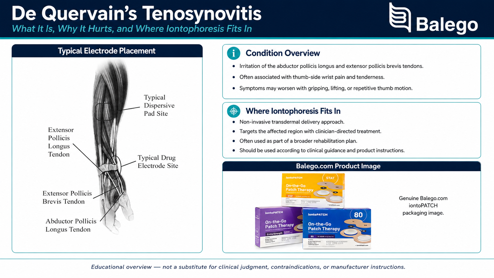
De Quervain’s Iontophoresis at a Glance
| Clinical question | Practical answer |
|---|---|
| What structures are involved? | The abductor pollicis longus and extensor pollicis brevis tendons within the first dorsal wrist compartment |
| Where is the pain usually located? | Near the radial styloid on the thumb side of the wrist |
| Is every case of radial wrist pain De Quervain’s? | No. Thumb CMC arthritis, intersection syndrome, superficial radial nerve irritation, fracture and other conditions may produce similar symptoms |
| Is iontophoresis the best-supported first treatment? | No. Comparative evidence most strongly supports corticosteroid injection combined with temporary thumb-spica immobilization |
| Where may iontophoresis fit? | As a prescribed, noninvasive adjunct when symptoms are localized and treatment is intended to improve participation in rehabilitation |
| Where is the drug electrode placed? | Over the clinically identified first dorsal compartment target—not automatically on the tip of the radial styloid |
| What should determine whether treatment continues? | Improved function, load tolerance and clinical findings, together with an acceptable skin response |
What Is De Quervain’s Tenosynovitis?
The first dorsal compartment lies along the thumb side of the wrist near the distal radius. It normally contains:
- Abductor pollicis longus, or APL: assists with moving the thumb away from the hand
- Extensor pollicis brevis, or EPB: assists with thumb extension
In De Quervain’s, thickening and reduced space around these tendons can make tendon gliding painful. Symptoms are often provoked by combined gripping, thumb movement, and wrist deviation.
Although the condition is frequently called “tendonitis,” it is better understood as a stenosing tenosynovial disorder rather than simply an acute inflammatory tendon injury.
Why anatomy matters
The APL and EPB do not always travel through one undivided tunnel. A septum may separate the EPB into its own subcompartment, and multiple APL tendon slips are common. These variations are particularly relevant when a patient has persistent symptoms or an incomplete response to an injection or surgical release. They do not mean that a larger iontophoresis electrode will necessarily deliver medication into every subcompartment.
Common Symptom Pattern
Patients commonly report:
- Pain over or slightly proximal to the radial styloid
- Pain with gripping, pinching or lifting
- Discomfort when moving the thumb away from the palm
- Pain during wrist deviation toward the little-finger side
- Difficulty opening jars or turning keys
- Pain while lifting an infant beneath the arms
- Local swelling or thickening
- Symptoms that may extend into the thumb or radial forearm
- Reduced confidence using the involved hand
Pregnancy and the postpartum period are recognized clinical contexts for De Quervain’s. Repetitive infant lifting may contribute mechanically, but pregnancy-related cases should not be reduced to “overuse” alone; hormonal, fluid, and gestational factors may also be relevant. Population-based and retrospective research supports an association between pregnancy or postpartum status and De Quervain’s.
Not All Thumb-Side Wrist Pain Is De Quervain’s
A focal diagnosis is essential before placing a medication electrode.
| Possible condition | Findings that may distinguish it from De Quervain’s |
|---|---|
| Thumb carpometacarpal osteoarthritis | Pain is centered closer to the base of the thumb; compression or grind testing may reproduce symptoms |
| Intersection syndrome | Pain and crepitus are generally located farther up the dorsal-radial forearm rather than directly near the radial styloid |
| Superficial radial nerve irritation | Burning, tingling, hypersensitivity or altered sensation may predominate |
| Scaphoid fracture or other wrist fracture | Trauma, snuffbox tenderness, swelling or pain with axial loading may be present |
| Scapholunate injury | Dorsal wrist pain, trauma history, instability or clicking may be more prominent |
| Trigger thumb | Catching or locking usually occurs near the volar thumb MCP region rather than the radial styloid |
| First-compartment ganglion | A discrete mass may be visible or palpable |
| Inflammatory arthritis | Multiple joints, bilateral symptoms, prolonged stiffness or systemic features may be present |
| Cervical or peripheral nerve referral | Symptoms may include paresthesia, weakness or a response to neck or nerve positioning |
| Infection | Increasing warmth, redness, swelling, fever or systemic illness requires medical evaluation |
When to Refer Before Beginning a Routine Protocol
Stop the routine modality pathway and arrange an appropriate medical assessment when findings suggest the following:
- Recent trauma with possible fracture
- Marked anatomical snuffbox tenderness
- Progressive swelling, redness or warmth
- Fever or suspected infection
- New or progressive neurological loss
- Significant vascular change
- An unexplained mass
- Severe pain that is not mechanically reproducible
- A sudden loss of thumb or wrist function
- Symptoms inconsistent with a first dorsal compartment disorder
- Medication allergy or another contraindication to the prescribed drug
Imaging is not required in every straightforward presentation. Ultrasound may be useful when the diagnosis is uncertain, symptoms persist despite appropriate treatment, or the clinician needs to evaluate first-compartment anatomy and possible septation. Ultrasound has demonstrated the ability to identify anatomical variations when compared with surgical findings.
De Quervain’s Examination Checklist
1. Clarify the history
Document:
- Exact symptom location
- Gradual or traumatic onset
- Duration and progression
- Dominant hand
- Occupational and household demands
- Infant-care responsibilities
- Tool, phone or computer use
- Sports or hobbies involving gripping
- Pain during and after activity
- Morning symptoms
- Previous wrist or thumb injury
- Prior splint, medication, therapy or injection response
- Pregnancy or postpartum status when relevant
- Medical conditions, medications and allergies
- Patient goals
Avoid assuming that any one activity “caused” the condition. The relationship between repetitive work and De Quervain’s is complex, and symptoms may reflect several interacting biological and mechanical factors.
2. Inspect the wrist and hand
Compare sides for:
- Local swelling
- Skin color
- Warmth
- Visible thickening
- A palpable mass
- Guarded thumb posture
- Scar tissue
- Brace-related skin irritation
- Skin suitability for electrode placement
3. Palpate the first dorsal compartment
Palpate along the APL and EPB path near and slightly proximal to the radial styloid.
Record:
- Exact location of maximal tenderness
- Length and width of the symptomatic region
- Whether the patient’s familiar pain is reproduced
- Presence of swelling or crepitus
- Whether tenderness is located farther proximal, suggesting intersection syndrome
- Whether symptoms are predominantly sensory, suggesting nerve involvement
- Whether a medication electrode can maintain complete skin contact
4. Assess thumb and wrist motion
Evaluate active and, when appropriate, passive.
- Thumb abduction
- Thumb extension
- Thumb flexion and opposition
- Wrist flexion and extension
- Radial deviation
- Ulnar deviation
- Forearm pronation and supination
Document pain location and movement quality—not only the number of degrees.
5. Assess strength and load tolerance
Depending on irritability, assess the following:
- Thumb abduction
- Thumb extension
- Grip
- Key pinch
- Three-jaw-chuck pinch
- Wrist radial deviation
- Functional lifting
- Jar-opening or twisting simulation
- Tool-holding tolerance
Grip and pinch testing may be deferred or modified when the patient is highly irritable.
Finkelstein’s Test Is Not the Same as Eichhoff’s Maneuver
These tests are frequently confused.
Eichhoff’s maneuver
The patient places the thumb inside the fist, and the wrist is then deviated toward the little-finger side.
This maneuver can place substantial stress on several radial wrist structures and may produce pain even in people without De Quervain’s.
Finkelstein’s test
In the classically described method, the examiner controls the thumb and gradually moves the hand or wrist into ulnar deviation.
A study of asymptomatic wrists found that Finkelstein’s test caused fewer false-positive responses and less discomfort than Eichhoff’s maneuver. However, because that study examined asymptomatic participants rather than using a definitive diagnostic reference standard in symptomatic patients, neither test should be treated as independently diagnostic.
Practical testing principle
A provocation test is most meaningful when it reproduces the patient’s recognizable first-compartment pain, not merely generalized discomfort from an aggressive wrist stretch.
Interpret test findings together with:
- Symptom location
- Palpation
- Movement response
- Strength
- Functional limitations
- Differential diagnosis
- Response to temporary load modification
What Is the Best-Supported Treatment?
A 2023 systematic review and network meta-analysis included 30 randomized studies and 1,663 patients. It found that corticosteroid injection combined with thumb-spica immobilization was associated with better pain and functional outcomes than several alternative strategies. The authors proposed corticosteroid injection plus three to four weeks of thumb-spica immobilization as a first-line approach. Ultrasound-guided injection was also associated with greater pain reduction than conventional injection in the included evidence.
That finding does not mean every patient must receive an injection. Treatment decisions may also depend on:
- Symptom severity and duration
- Pregnancy or lactation considerations
- Medical history
- Medication risks
- Patient preferences
- Access to injection services
- Prior response
- Occupational requirements
- Clinical examination findings
Where Does Iontophoresis Fit?
Iontophoresis uses controlled direct current to support the delivery of an appropriately ionized medication through intact skin. The full explanation of current, medication polarity, and mA-min dosing is already available in our Iontophoresis Frequently Asked Questions and should not be duplicated on this diagnosis-specific page.
Iontophoresis may be considered when:
- The diagnosis is reasonably clear
- Symptoms are focal and superficial
- The skin is intact
- A prescriber has selected an appropriate medication
- The patient or clinician prefers a non-needle delivery approach
- Pain is preventing participation in therapeutic exercise or daily activity
- The treatment target can be covered without placing the electrode directly over an unsuitable bony contour
- A defined reassessment plan is established
Iontophoresis should not be presented as
- A proven equivalent to corticosteroid injection
- A treatment that mechanically opens the first dorsal compartment
- A method of correcting a septum or anatomical variation
- A replacement for activity modification or rehabilitation
- A guaranteed way to prevent surgery
- Appropriate for undiagnosed radial wrist pain
- A patient-directed medication treatment without professional oversight
What Does the Research Say About De Quervain’s Iontophoresis?
| Evidence source | Main finding | Important limitation |
|---|---|---|
| Retrospective hand-therapy outcome study | Dexamethasone iontophoresis was associated with improved functional outcomes | Level IV retrospective evidence cannot establish that iontophoresis caused the improvement |
| 2023 De Quervain network meta-analysis | The best comparative evidence favored corticosteroid injection with temporary thumb-spica immobilization | Iontophoresis did not have comparable high-quality evidence |
| 2025 conservative-treatment review | The relative effectiveness of conservative interventions remained uncertain across a varied evidence base | Heterogeneous treatments and study quality limit direct comparisons |
| 2026 three-patient tendinopathy case series | One De Quervain case improved during multimodal therapy that included dexamethasone iontophoresis | A case series cannot separate the effect of iontophoresis from splinting, exercise or natural recovery |
The De Quervain-specific iontophoresis literature is therefore suggestive, not conclusive. The retrospective study reported a functional association, but the evidence level was low. A recent case series also reported improvement during multimodal care, but it included only one De Quervain patient and cannot determine which component produced the result.
Comparing Treatment Options
| Treatment | Primary purpose | Relative evidence | Advantages | Limitations |
|---|---|---|---|---|
| Activity and technique modification | Reduce repeated aggravation while maintaining necessary hand use | Common component of conservative care | Low risk and immediately applicable | May be insufficient alone |
| Thumb-spica orthosis | Temporarily limits provocative thumb and wrist movement | Best supported when combined with corticosteroid injection | Noninvasive and useful during high-load activities | Poor fit, excessive immobilization or inconsistent use can limit benefit |
| Therapeutic exercise | Restore motion, strength and task tolerance | A common rehabilitation component, but protocols vary | Addresses function and capacity | Requires appropriate dosage and progression |
| Corticosteroid injection | Deliver medication directly into the first dorsal compartment | Strongest comparative nonsurgical evidence | May produce meaningful pain and functional improvement | Invasive medication and procedural risks require medical review |
| Iontophoresis | Deliver prescribed ionized medication through intact skin | Limited De Quervain-specific evidence | Non-needle, localized and compatible with rehabilitation workflows | It does not mechanically release the compartment; skin response and electrode fit matter |
| Surgical release | Decompress the first dorsal compartment | Used for persistent or refractory cases | Addresses mechanical stenosis directly | Operative risks, recovery and scar considerations |
Step-by-Step De Quervain’s Iontophoresis Protocol
Step 1: Confirm the clinical target
Identify the first dorsal compartment and confirm that palpation or controlled loading reproduces the patient’s familiar symptoms.
Do not select the treatment site solely from a generic anatomical illustration.
Document whether the tenderness is:
- Directly adjacent to the radial styloid
- Slightly proximal to the styloid
- Distributed along the APL and EPB path
- Farther proximal, raising concern for intersection syndrome
- Predominantly at the thumb CMC joint
- Associated with sensory symptoms
Step 2: Define the treatment objective
Examples include:
- Improve tolerance for thumb-spica weaning
- Reduce pain that prevents gentle thumb motion
- Improve ability to participate in strengthening
- Improve tolerance for infant-care techniques
- Reduce pain during a defined work task
- Improve grip or pinch tolerance
“Reduce inflammation” is not a complete functional objective by itself.
Step 3: Verify the medication order
Confirm:
- Medication name
- Concentration
- Medication polarity
- Prescribed dose
- Treatment frequency
- Medication allergies
- Patient-specific precautions
- Compatibility with the selected electrode or patch
Medication is not included with iontophoresis controllers, electrodes, or patches.
Use our Iontophoresis Electrode Directions for Use for the current polarity table, filling instructions, dose limits, warnings, and skin safety information.
Step 4: Inspect the skin and remove interfering items
Inspect both the medication-electrode and return-electrode sites for:
- Cuts
- Abrasions
- Dermatitis
- Recent scar tissue
- Altered sensation
- Brace-related irritation
- Jewelry contact
- Lotion or oil
- Excessive hair
- Prior electrode reactions
Remove watches, bracelets, and jewelry that could contact either electrode or lead.
Step 5: Choose an electrode that matches the wrist contour
The first dorsal compartment is a narrow, mobile, and curved treatment region. A smaller or flexible electrode may maintain better contact than a large flat electrode.
The selected drug matrix should cover the focal target without the following issues:
- Wrapping excessively around the wrist
- Folding
- Bridging the anatomical snuffbox
- Entering a deep skin crease
- Extending onto irritated skin
- Placing the entire drug chamber over the radial styloid tip
Step 6: Position the medication electrode
Place the active drug-delivery area lengthwise over the clinically identified APL and EPB region.
A practical target is often slightly proximal to the radial styloid, where the electrode can cover the first compartment while maintaining full contact. Exact positioning varies according to anatomy, symptom location, and electrode shape.
Avoid:
- Placing the reservoir indiscriminately in the anatomical snuffbox
- Centering it over an unrelated thumb CMC target
- Allowing the adhesive to wrinkle
- Applying pressure over a bony prominence
- Covering areas with altered sensation
- Taping tightly around the wrist
Step 7: Position the return electrode
For a controller-based treatment, place the return electrode over an appropriate area of intact forearm tissue according to the electrode and controller instructions.
Confirm:
- Complete contact
- Adequate distance from the active electrode
- No interference from clothing
- No compression from an orthosis
- No lead-wire tension
- No jewelry between or near the electrodes
- Visibility or accessibility for monitoring
Step 8: Position and support the hand
Support the forearm with the wrist in a comfortable neutral or near-neutral position and the thumb relaxed.
During a controller-based session, avoid repetition of:
- Thumb flexion and extension
- Wrist flexion and extension
- Forceful radial or ulnar deviation
- Gripping
- Pinching
- Lead-wire pulling
Movement can lift an electrode edge, disrupt current, or concentrate pressure.
Step 9: Deliver the prescribed dose
The source protocol used a 40 mA-min dose, but this value should not be presented as universal.
With the ActivaDose II Iontophoresis Dose Controller, the clinician selects the prescribed dose and current, and the controller calculates the required treatment time. Its adjustable range extends to 80 mA-min, but the permitted treatment depends on medication polarity, electrode limits, skin response, and the prescription.
Current electrode directions state the following:
- Negative-polarity delivery must not exceed the listed negative-polarity dose and current limits
- Positive-polarity delivery has a lower listed maximum dose
- Pain requires immediate attention
- Interrupted contact or an electrode-reject warning must be corrected before continuing
Follow the exact instructions supplied with the product being used rather than transferring limits from one electrode system to another.
Step 10: Monitor the patient throughout treatment
Ask the patient to report:
- Burning
- Sharp pain
- Increasing stinging
- Strong itching
- New numbness or tingling
- Electrode movement
- Lead tension
- Pressure from the table, clothing or brace
Pause and inspect the skin when discomfort increases unexpectedly. Do not simply lower the current and continue without investigating the cause.
Step 11: Inspect, document and functionally reassess
After treatment, document:
- Medication and concentration
- Polarity
- Dose
- Current
- Treatment time
- Electrode or patch model
- Exact anatomical placement
- Patient sensation
- Skin appearance
- Immediate change in tenderness
- Change in the selected movement or functional task
A transient skin response can occur during direct-current treatment. Blistering, persistent pain, pronounced irritation, or an unusual rash requires appropriate follow-up.
Comparing Balego Iontophoresis Products for the Radial Wrist
This table focuses on De Quervain-specific placement and workflow. Full specifications remain on the linked Balego product pages.
| Balego option | Potential radial-wrist advantage | Important limitation | Practical fit |
|---|---|---|---|
| ActivaDose II Controller with Trivarion electrodes | Flexible, form-fitting electrode choices can help maintain contact on a small or curved wrist region | Requires lead wires, a controller and monitored treatment positioning | Clinics prioritizing adjustable current, dose control and direct observation |
| ActivaDose II with Ionto4 electrodes | Small-square and butterfly options provide different footprints and reservoir capacities | A square electrode may be less conforming when centered too close to the radial styloid | Clinics wanting multiple electrode sizes in a standardized disposable kit |
| ActivaDose II with Balego buffered electrode kits | Includes active and buffered return electrodes with several size and shape choices | Electrode size must match the small treatment target; larger is not automatically better | Controller-based clinics wanting a buffered return-electrode workflow |
| IontoPATCH wearable patches | The self-contained butterfly format eliminates external wires and offers several dose/wear-time options | Wrist motion, sleeves, handwashing and splint pressure can affect adhesion or comfort | Patients who can protect the patch throughout the prescribed wear period |
| ACTIVApatch wearable patches | Self-contained options include shorter and extended treatment windows | Medication polarity, patch footprint and movement tolerance must match the application | Clinics seeking wireless delivery with several scheduling options |
ActivaDose II supports adjustable controller-based delivery. Trivarion is designed for curved or uneven anatomical regions and is available in small, medium, butterfly, and large configurations. Ionto4 also provides small-square and butterfly options. IontoPATCH and ACTIVApatch offer wireless workflows but require careful planning for a high-movement wrist location.
For a complete wearable-system comparison, link to our IontoPatch vs. ACTIVApatch Guide rather than duplicating every dose, reservoir, and treatment-time specification here.
Can a Thumb-Spica Orthosis Be Worn Over a Patch?
Do not automatically place a rigid or tight orthosis over an active iontophoresis patch.
An orthosis may:
- Compress the medication reservoir
- Lift or roll the adhesive edge
- Trap perspiration
- Increase friction
- Concentrate pressure over the radial styloid
- Prevent inspection of the skin
- Change electrode contact during movement
When both treatments are prescribed, plan their timing so that the orthosis and patch do not interfere. Follow the patch instructions and clinician’s direction regarding compression, wear, and removal.
Rehabilitation After Symptoms Become More Tolerable
Iontophoresis should help restore useful hand function.
Phase 1: Reduce unnecessary irritation
Modify how the patient performs aggravating tasks rather than simply advising “rest.”
Examples include:
- Lift an infant with the wrists straighter and the forearms supporting more of the load
- Use both hands for pans, containers and heavy objects
- Avoid repeatedly lifting with the thumbs spread wide
- Enlarge tool or utensil handles when appropriate
- Reduce forceful pinching
- Alternate hands and tasks
- Use voice input or different phone positioning
- Take brief recovery periods during repetitive work
- Avoid repeatedly retesting the painful maneuver
Phase 2: Restore comfortable motion
When irritability permits, use controlled
- Thumb flexion and extension
- Thumb abduction and opposition
- Wrist flexion and extension
- Forearm rotation
- Tendon-gliding movement
Do not force an aggressive Finkelstein- or Eichhoff-type stretch as a routine exercise.
Phase 3: Restore strength and capacity
Progress gradually through:
- Pain-limited isometric thumb abduction
- Thumb-extension resistance
- Radial-deviation resistance
- Grip training
- Pinch training
- Object manipulation
- Controlled lifting
- Task-specific endurance
Loading should reproduce no more than an acceptable level of symptoms and should not cause a disproportionate increase afterward.
Phase 4: Return to the required task
Progress the activity that matters to the patient:
- Infant care
- Food preparation
- Scissor use
- Computer work
- Tool handling
- Racquet or stick sport
- Gardening
- Musical performance
- Clinical or laboratory work
The goal is not simply a negative provocation test. It is a restored capacity for meaningful hand use.
Reassessment Checklist
Repeat the measures established at the initial examination.
Symptoms
- Local tenderness
- Pain at rest
- Pain during thumb motion
- Pain during wrist deviation
- Swelling
- Sensory symptoms
- Skin response
Motion and strength
- Thumb abduction
- Thumb extension
- Wrist movement
- Grip
- Pinch
- Task-specific force tolerance
Function
- Lifting an infant
- Opening a jar
- Gripping a pan
- Using scissors
- Holding a phone
- Performing work tasks
- Returning to sport or hobbies
- QuickDASH or Patient-Specific Functional Scale score
Reasons to modify or stop the plan
- No meaningful functional improvement during the defined treatment trial
- Increasing swelling or pain
- New sensory symptoms
- Repeated skin irritation
- Inability to maintain electrode contact
- Symptoms localizing away from the first dorsal compartment
- Findings suggesting thumb CMC arthritis, intersection syndrome or nerve involvement
- Dependence on passive treatment without progression to functional rehabilitation
- Concern for an alternative diagnosis
Suggested Treatment-Plan Comparison
| Patient presentation | Possible priority |
|---|---|
| Mild, recent symptoms with a clear task-related trigger | Education, technique modification and selective orthosis use |
| Symptoms limiting daily activity despite initial modification | Structured hand therapy and reassessment of diagnosis |
| Localized symptoms with an appropriate medication order and a preference for non-needle delivery | Consider iontophoresis as an adjunct |
| Persistent or functionally significant symptoms | Discuss the evidence for corticosteroid injection with temporary thumb-spica immobilization |
| Recurrent symptoms or incomplete response after appropriate nonsurgical care | Evaluate anatomy, diagnosis and need for specialist referral |
| Refractory mechanical stenosis | Surgical consultation may be appropriate |
Documentation Template
Clinical diagnosis or working classification: De Quervain’s tenosynovitis involving the right/left first dorsal compartment.
Differential diagnoses considered: __________.
Focal treatment target: Tenderness is located __________ relative to the radial styloid.
Functional limitation: Pain limits __________.
Baseline measure: QuickDASH/PSFS/grip/pinch/task result __________.
Reason iontophoresis was selected: Prescribed localized medication delivery intended to improve tolerance for __________.
Medication order: Medication __________; concentration __________; polarity __________.
Delivery system: Controller/patch __________; electrode size or model __________.
Placement: Medication electrode __________; return electrode __________.
Treatment details: Dose __________ mA-min, current __________ mA, and time ___________.
Patient response: Sensation __________; skin response __________.
Post-treatment functional finding: __________.
Rehabilitation plan: __________.
Reassessment date or criterion: __________.
For payer-oriented information, direct clinicians to our Iontophoresis Reimbursement Codes and Documentation Guide.
Frequently Asked Questions
Can iontophoresis help De Quervain’s tenosynovitis?
It may help selected patients when a clinician identifies a focal first-compartment target and a prescriber orders an appropriate ionized medication. The evidence is limited and substantially weaker than the evidence supporting corticosteroid injection combined with temporary thumb-spica immobilization.
Is iontophoresis a substitute for a corticosteroid injection?
It should not be described as an equivalent substitute. It is a different method of medication delivery with a different evidence base. It may be considered when clinically appropriate, but treatment choice requires professional evaluation and patient-specific decision-making.
Where should the medication electrode be placed?
Place the medication reservoir over the focal APL and EPB treatment target identified during examination, commonly near or slightly proximal to the radial styloid. Avoid simply centering it over the bony tip or in the anatomical snuffbox without confirming the actual symptomatic structure.
Should the electrode cover both APL and EPB tendons?
The electrode should cover the clinically identified first-compartment region as its size and shape permit. Anatomical septation can separate the EPB, so surface placement should not be described as guaranteeing medication delivery equally throughout every internal subcompartment.
Is dexamethasone included with the patch or electrodes?
No. Medication is supplied separately and must be prescribed or otherwise ordered by an appropriately licensed professional.
Is dexamethasone always delivered from the negative electrode?
Dexamethasone sodium phosphate is commonly treated as negatively charged, but clinicians must verify the exact medication formulation, polarity, and product instructions before every application.
Is 40 mA-min always the correct dose?
No. The source protocol uses 40 mA-min, but the dose depends on the medication order, polarity, electrode or patch limits, controller, skin condition, and patient response.
How many treatments should be performed?
There is no universal number. The original protocol described three to six sessions every other day, but that schedule should not be applied automatically. Establish a defined treatment trial and continue only when reassessment demonstrates meaningful benefit.
Can a patient move the wrist during treatment?
Movement should be minimized during controller-based delivery because it can disturb electrode contact and create lead-wire tension. A wearable patch may permit mobility, but repeated thumb and wrist movement can still affect adhesion and skin comfort.
Can the patient wear a thumb-spica brace over an iontophoresis patch?
Not automatically. A brace may compress or displace the patch. Coordinate the timing of the treatments and follow the device instructions and clinician’s directions.
Is Finkelstein’s test enough to diagnose De Quervain’s?
No. A provocative test supports the examination but does not replace history, palpation, motion, strength testing, and consideration of other causes of radial wrist pain.
When should imaging be considered?
Imaging may be useful when the diagnosis remains uncertain, trauma is present, another disorder is suspected or symptoms persist despite an appropriate treatment trial. Ultrasound can also help identify first-compartment septation and other anatomical variations.
Clinical Takeaway
A useful De Quervain’s protocol begins by confirming that the pain truly originates from the first dorsal compartment.
The clinician should localize the symptoms, distinguish Finkelstein’s test from Eichhoff’s maneuver, screen for competing causes of radial wrist pain, and establish measurable functional limitations. Current comparative evidence most strongly supports corticosteroid injection combined with temporary thumb-spica immobilization. Iontophoresis has a smaller and lower-quality evidence base, but it may be considered as a clinician-directed, non-needle adjunct when the target is localized and a suitable medication has been prescribed.
The treatment should have a clear purpose: helping the patient resume motion, strengthening, infant care, work, sport, or another meaningful activity. When function does not improve, repeating the same passive treatment is less appropriate than reconsidering the diagnosis and overall plan.
Explore our complete selection of iontophoresis patches, electrodes, and dose controllers, or browse additional iontophoresis evaluation and treatment protocols.


