



body{font-family:system-ui,-apple-system,Segoe UI,Roboto,Ubuntu,Cantarell,Noto Sans,sans-serif;line-height:1.6;margin:0;padding:32px;background:#fff;color:#111} h1,h2{color:#0f172a;margin-bottom:.4em} h1{font-size:clamp(1.6rem,2.2vw,2.2rem)} h2{font-size:clamp(1.2rem,1.9vw,1.6rem);margin-top:1.4em} h3{margin:.9em 0 .3em} ul,ol{margin:.5em 0 .9em 1.2em} table{width:100%;border-collapse:collapse;margin:1em 0;background:#fff;border:1px solid #e5e7eb} th,td{padding:10px 12px;border-bottom:1px solid #e5e7eb;vertical-align:top} th{background:#eef6ff;text-align:left} .card{background:#f8fafc;border:1px solid #e5e7eb;border-radius:12px;padding:14px;margin:.8em 0} .tiny{font-size:.9rem;color:#475569} footer{margin-top:24px;font-size:.9rem;color:#475569}
Golfer’s elbow, traditionally called medial epicondylitis, is pain arising from the common flexor-pronator tendon near the inside of the elbow. It is associated with repetitive or excessive gripping, wrist flexion, forearm pronation, throwing, lifting, and tool use.
The newer term "medial elbow tendinopathy" is often more anatomically accurate. Persistent cases commonly involve tendon disorganization and degeneration rather than a purely inflammatory process. The condition can affect golfers, but it is also seen in throwing athletes, racquet-sport participants, tradespeople, healthcare workers, weightlifters, and anyone whose work or recreation repeatedly loads the wrist flexors and forearm pronators.
Iontophoresis may be considered when:
- Symptoms are localized near the common flexor-pronator origin.
- The diagnosis has been reasonably confirmed.
- The skin is intact.
- A prescriber has selected an appropriate ionized medication.
- Medication polarity has been verified.
- The patient is suitable for direct-current treatment.
- Short-term symptom control has a defined rehabilitation purpose.
- Progress will be measured objectively.
Iontophoresis should be positioned as a clinician-directed medication-delivery adjunct, not as a cure, tendon-repair procedure, or proven replacement for injection.
For general medication, polarity, filling, dose limit, and device instructions, use Balego’s Iontophoresis Electrode Directions for Use. This protocol concentrates on the medial elbow rather than repeating that material.
Clinical scope: This guide is intended for licensed healthcare professionals. It does not prescribe medication, concentration, polarity, current, electrical dose, treatment frequency, or total number of applications.
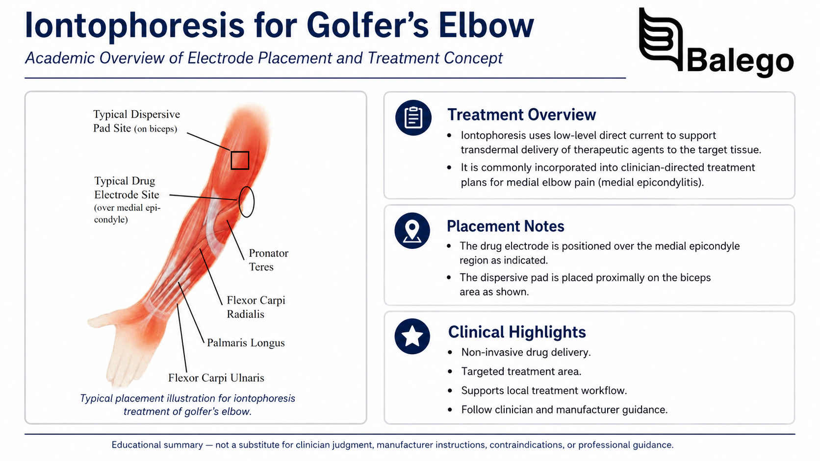
Golfer’s Elbow Iontophoresis: Quick Clinical Summary
| Clinical question | Practical answer |
|---|---|
| What tissue is usually involved? | The common flexor-pronator tendon at or just distal to the medial epicondyle |
| Which muscles contribute to the tendon? | Pronator teres, flexor carpi radialis, palmaris longus, flexor digitorum superficialis, and flexor carpi ulnaris |
| What commonly reproduces symptoms? | Resisted wrist flexion, forearm pronation, gripping, and passive wrist-extension stretch |
| Where is the medication electrode positioned? | Over the most tender portion of the common flexor-pronator origin |
| What nearby structure requires special attention? | The ulnar nerve behind the medial epicondyle |
| Should the drug electrode cover the cubital tunnel? | Generally no; avoid placing concentrated current directly over the symptomatic ulnar nerve |
| Where does the return electrode go? | Over intact nearby muscle tissue according to the electrode-system instructions |
| Is 40 mA·min always required? | No, it is a frequently studied dose, not a universal prescription |
| Is six treatments the standard course? | No; six treatments came from an older combined medial/lateral epicondylitis trial |
| What should accompany treatment? | Load modification, progressive tendon loading, grip rehabilitation, proximal strengthening, and activity retraining |
| When is referral important? | Trauma, instability, neurological symptoms, youth throwing injury, marked weakness, or failure to improve |
Medial Elbow Anatomy and Why It Matters
The medial epicondyle is the bony prominence on the inside of the elbow. The common flexor tendon attaches in this region and connects several muscles responsible for wrist flexion, finger flexion, and forearm pronation.
| Muscle | Principal contribution relevant to golfer’s elbow |
|---|---|
| Pronator teres | Turns the palm downward and assists elbow flexion |
| Flexor carpi radialis | Flexes the wrist and moves it toward the thumb side |
| Palmaris longus | Assists wrist flexion when present |
| Flexor digitorum superficialis | Flexes the fingers and contributes to forceful gripping |
| Flexor carpi ulnaris | Flexes the wrist and moves it toward the little-finger side |
In medial epicondylitis, clinicians frequently emphasize the pronator teres and flexor carpi radialis, although symptoms may also involve the broader flexor-pronator origin.
The ulnar nerve is immediately nearby
The ulnar nerve passes behind the medial epicondyle through the cubital-tunnel region. This creates two important clinical implications:
- Medial epicondylitis may coexist with ulnar-nerve irritation.
- Not every case of medial elbow pain is a tendon disorder.
Numbness, tingling, burning, or altered sensation in the ring and little fingers should prompt a more complete ulnar-nerve assessment. Weakness or wasting of ulnar-innervated hand muscles requires medical evaluation rather than simply repositioning an iontophoresis electrode.
Typical Golfer’s Elbow Symptoms
Common features include:
- Gradual pain at the inside of the elbow
- Tenderness over or just below the medial epicondyle
- Pain during forceful gripping
- Pain with wrist flexion
- Pain with forearm pronation
- Reduced grip tolerance
- Pain when lifting with the palm up
- Symptoms during golf, throwing, climbing, weightlifting, or tool use
- Stiffness after activity
- Difficulty opening containers or carrying objects
Common Causes and Loading Patterns
Golfer’s elbow generally reflects repeated tendon loading that exceeds current tissue capacity.
| Loading pattern | Examples |
|---|---|
| Repeated gripping | Tools, weights, climbing holds, racquets, golf clubs |
| Wrist flexion under load | Lifting, curls, manual work, repetitive pulling |
| Repeated pronation | Screwdriver use, throwing, racquet or bat movement |
| High-force finger flexion | Carrying, gripping handles, occupational pinch and grasp |
| Throwing stress | Baseball, softball, javelin, football |
| Sudden training increase | More repetitions, heavier weights, increased practice volume |
| Poor technique | Late golf impact, excessive wrist action, throwing-mechanics errors |
| Equipment mismatch | Incorrect grip size, heavy tool, unsuitable racquet or club |
| Insufficient recovery | Repeated loading without adequate adaptation |
| Direct trauma | Impact to the medial elbow or acute flexor-pronator strain |
A treatment plan should identify the provoking load rather than focusing only on where the elbow hurts.
What Does the Evidence Say About Iontophoresis?
The principal epicondylitis trial
A randomized, double-blinded, placebo-controlled study evaluated dexamethasone sodium phosphate iontophoresis in 199 patients with acute medial or lateral epicondylitis.
The research protocol used:
- Symptoms generally lasting three months or less
- Six treatments
- Treatments spaced one to three days apart
- Completion within 15 days
- A 40 mA·min electrical dose
- Dexamethasone sodium phosphate 0.4% or saline placebo
At two-day follow-up, the dexamethasone group had greater pain improvement and more favorable investigator ratings. At one month, differences between active and placebo groups were not statistically significant for the principal outcomes. The researchers therefore supported short-term symptom reduction—not a durable or medial-elbow-specific superiority claim.
Important limitations
| Limitation | Why it matters |
|---|---|
| Medial and lateral cases were pooled | The study did not establish a separate medial-epicondylitis effect size |
| Participants had relatively acute symptoms | Results may not transfer to long-standing degenerative tendinopathy |
| The benefit was strongest shortly after treatment | The advantage was not statistically significant at one month |
| One medication and concentration were studied | The findings do not validate every medication formulation |
| The product workflow was controller-based. | Results cannot automatically be transferred to every wearable patch |
| Electrode-site reactions were common | Skin monitoring remains essential |
| The study had commercial product funding | Funding context should be disclosed when interpreting results |
The trial also stated that the ideal number and spacing of iontophoresis treatments had historically been based more on clinical experience than robust comparative research.
Responsible evidence statement
Iontophoresis may help reduce symptoms over the short term in selected patients with acute epicondylitis. Current evidence does not establish it as
- A stand-alone tendon-healing treatment
- Superior to progressive rehabilitation
- Equivalent to injection
- Proven specifically for chronic medial epicondylitis
- A guaranteed way to prevent surgery
- Appropriate for every source of medial elbow pain
Evaluate the Patient Before Treating the Elbow
Subjective history
| Question | Why it matters |
|---|---|
| Where is the pain located? | Distinguishes common flexor origin from cubital tunnel, UCL, joint, or referred pain |
| How did it begin? | Separates gradual overload from acute strain, fracture, or ligament injury |
| Which movements provoke it? | Identifies grip, flexion, pronation, throwing, lifting, or occupational load |
| Is pain present during or after activity? | Helps assess irritability and load response |
| Is there numbness or tingling? | Screens for ulnar neuropathy |
| Is the elbow unstable, or does it “give way”? | Raises concern for ligament injury |
| Was there a pop, sudden pull, or bruising? | May indicate acute tendon or ligament damage |
| Is the patient a throwing athlete? | Requires attention to valgus load and the UCL |
| Is the patient skeletally immature? | Medial epicondyle apophysitis or avulsion may mimic golfer’s elbow |
| What work or sport must be resumed? | Defines functional goals and return criteria |
| What treatments have already been tried? | Prevents indefinite repetition of ineffective care |
| Which medications and allergies are present? | Required before iontophoresis |
| Is there skin or adhesive sensitivity? | Affects electrode or patch selection |
Observation
Assess:
- Swelling
- Bruising
- Redness or warmth
- Skin integrity
- Surgical scars
- Carrying angle
- Protective arm posture
- Forearm-muscle asymmetry
- Elbow extension during functional use
- Throwing or lifting mechanics when relevant
Palpation
Palpate systematically:
- Medial epicondyle
- Common flexor-pronator tendon
- Pronator teres origin
- Flexor carpi radialis region
- Ulnar nerve and cubital tunnel
- Ulnar collateral ligament
- Medial joint line
- Olecranon
- Distal biceps and brachialis when indicated
The medication target should be the reproducible tendon pain—not simply the most prominent bone.
Range of motion
Measure:
- Elbow flexion and extension
- Forearm pronation and supination
- Wrist flexion and extension
- Radial and ulnar deviation when relevant
- Cervical and shoulder movement if symptoms suggest a regional contribution
Limited motion is not always a defining feature of golfer’s elbow. Marked joint restriction should prompt consideration of arthritis, trauma, effusion, or another elbow disorder.
Strength and function
Assess according to symptom irritability:
- Wrist flexion strength
- Forearm pronation strength
- Pain-free grip strength
- Finger-flexion tolerance
- Elbow flexion and extension
- Shoulder and scapular strength
- Lifting tolerance
- Carrying tolerance
- Work-tool use
- Golf, racquet, climbing, or throwing tasks
Pain-free grip strength is often more useful than maximal grip because it records the force tolerated before symptoms appear.
Provocation Tests
No single test should be treated as diagnostic in isolation.
| Test | Basic method | Positive response |
|---|---|---|
| Resisted wrist flexion | Elbow supported while the patient flexes the wrist against resistance | Familiar pain at the common flexor origin |
| Resisted forearm pronation | Patient turns the palm downward against resistance | Familiar medial epicondyle or proximal forearm pain |
| Combined flexion-pronation test | Wrist flexion and pronation resisted together | Reproduction of localized tendon symptoms |
| Passive wrist-extension stretch | Elbow extended with the forearm supinated and wrist/fingers extended | Stretch pain at the flexor-pronator origin |
| Grip test | Grip dynamometer or functional object | Medial elbow pain or reduced pain-free grip |
| Tinel test at cubital tunnel | Gentle percussion over the ulnar nerve | Tingling into the ring or little finger |
| Valgus-stress testing | Performed when ligament injury is suspected | Medial pain, laxity, or apprehension requiring further assessment |
The diagnosis is supported by localized tenderness and pain reproduced with resisted wrist flexion or pronation. A comprehensive examination remains necessary because the medial elbow contains the common flexor tendon, UCL, ulnar nerve, joint structures, and medial epicondyle within a small region.
Differential Diagnosis of Medial Elbow Pain
| Possible condition | Findings that may distinguish it |
|---|---|
| Ulnar neuropathy or cubital-tunnel syndrome | Tingling or numbness in the ring and little fingers, hand weakness, symptoms with sustained elbow flexion |
| Ulnar collateral ligament injury | Throwing history, valgus pain, instability, reduced throwing velocity |
| Flexor-pronator strain or tear | Acute onset, bruising, weakness, pain after a specific forceful event |
| Medial epicondyle apophysitis | Skeletally immature throwing athlete with medial-elbow pain |
| Medial epicondyle avulsion fracture | Acute trauma or throwing injury, swelling, marked tenderness, loss of function |
| Elbow osteoarthritis | Joint-line pain, stiffness, reduced range, mechanical symptoms |
| Cervical radiculopathy | Neck symptoms, radiating pain, reflex or sensory changes |
| Pronator syndrome | Median-nerve symptoms with proximal forearm pain |
| Ulnar-nerve instability | Snapping or subluxation behind the medial epicondyle |
| Snapping triceps | Medial snapping during elbow movement |
| Inflammatory or infectious condition | Marked warmth, redness, swelling, fever, or systemic illness |
| Referred shoulder or wrist pain | Symptoms reproduced outside the local elbow examination |
Ultrasound or MRI may be useful when the diagnosis is uncertain, a tear or ligament injury is suspected, symptoms are atypical, or conservative care is unsuccessful. Medial elbow ultrasound must differentiate tendon pathology from ulnar-nerve instability, UCL injury, snapping triceps, and joint disease.
Red Flags and Referral Criteria
Refer for medical evaluation before iontophoresis when there is:
- Acute trauma with a pop or sudden loss of strength
- Marked swelling or bruising
- Suspected tendon tear
- Valgus instability
- Suspected UCL injury
- Numbness, tingling, or progressive hand weakness
- Muscle wasting
- Inability to grip or use the arm normally
- Loss of elbow motion after trauma
- Fever, redness, drainage, or suspected infection
- Severe night or resting pain
- A palpable mass
- Symptoms in a child or adolescent throwing athlete
- Possible medial epicondyle apophyseal or avulsion injury
- An implanted device or medical condition prohibited by the iontophoresis labeling
- Uncertainty about the prescribed medication or polarity
Little League elbow and medial epicondyle avulsion injuries should not be treated as adult golfer’s elbow. Skeletally immature athletes require a separate diagnostic and treatment pathway.
When Iontophoresis May Fit
A clinician may consider iontophoresis when:
- Pain is localized to the superficial common flexor-pronator origin.
- Symptoms are mild to moderate.
- The presentation is nontraumatic.
- Neurologic symptoms are absent or separately evaluated.
- The elbow is stable.
- A prescriber has ordered an appropriate medication.
- The exact formulation and polarity are known.
- The skin is intact.
- An electrode can fit without covering or compressing the cubital tunnel.
- Symptoms limit participation in a progressive loading program.
- A defined reassessment point has been established.
When another approach may be more appropriate
| Finding | Clinical response |
|---|---|
| Predominant ring- or little-finger tingling | Evaluate the ulnar nerve |
| Throwing-related instability | Evaluate the UCL and throwing mechanics |
| Acute weakness or bruising | Rule out flexor-pronator tear |
| Child or adolescent athlete | Rule out apophyseal injury |
| Diffuse joint pain and stiffness | Assess intra-articular pathology |
| Skin breakdown near the target | Do not place the electrode over the area |
| No response to a defined trial | Reconsider diagnosis and treatment strategy |
| Longstanding degenerative symptoms | Emphasize progressive loading rather than repeated passive treatment |
| Inability to fit the patch safely | Select a smaller controller-based electrode or another treatment |
Golfer’s Elbow Iontophoresis Protocol Framework
Step 1: Confirm the medical order
Before opening an electrode or patch, verify:
- Medication name
- Exact chemical formulation
- Concentration
- Prescribed volume
- Ionic charge
- Required polarity
- Electrical dose
- Maximum current
- Treatment frequency
- Planned number of applications
- Medication allergies
- Treatment location
Medication is supplied separately from Balego’s iontophoresis controllers, electrodes, and wearable patches.
Use the Iontophoresis FAQ, Medication, Dose, and Polarity Guides when clarification is needed, but confirm the exact order with the prescriber or pharmacist.
Step 2: Record baseline outcomes
Useful measures include:
| Outcome | Baseline method |
|---|---|
| Pain at rest | 0–10 rating |
| Pain with gripping | 0–10 rating during a standardized grip |
| Pain-free grip | Dynamometer or repeatable functional task |
| Wrist-flexion tolerance | Resistance level before familiar pain |
| Pronation tolerance | Resistance level before familiar pain |
| Local tenderness | Standardized palpation site |
| Function | QuickDASH, Patient-Specific Functional Scale, or clinic-selected measure |
| Work tolerance | Time, repetitions, weight, or task duration |
| Sport tolerance | Swings, throws, climbs, or lifts before symptom increase |
| Neurologic status | Sensation and ulnar-nerve findings |
| Skin condition | Document both planned electrode sites |
Step 3: Identify and mark the tendon target
Locate the most reproducible tender point at the common flexor-pronator origin.
The target is typically:
- At or slightly distal and anterior to the medial epicondyle
- Within the common flexor-pronator tendon region
- Reproduced by resisted wrist flexion or pronation
- Separate from the ulnar-nerve groove behind the medial epicondyle
Mark the site before skin preparation.
Step 4: Choose an electrode system that fits the elbow
Consider:
- Size of the tender region
- Elbow contour
- Medication volume
- Prescribed dose
- Need for current adjustment
- Skin sensitivity
- Adhesive tolerance
- Patient schedule
- Whether the elbow will move repeatedly
- Proximity to the ulnar nerve
- Need for direct clinical observation
A flexible or butterfly-shaped controller electrode may conform better to the curved medial elbow than a large flat patch.
Step 5: Prepare the skin according to the product
Do not use one skin-preparation rule for every iontophoresis system.
- Some controller-electrode kits include alcohol preparation pads.
- Wearable patches may specify soap-and-water preparation.
- The selected controller, electrode, and patch instructions remain controlling.
General principles include:
- Use intact skin.
- Remove lotion, oil, and heavy contamination.
- Allow the site to dry fully.
- Clip excessive hair when necessary.
- Do not shave immediately before treatment.
- Avoid abrasions, eczema, wounds, recent injections, and irritated skin.
- Inspect both the medication and return-electrode sites.
Review Iontophoresis Electrode Directions for Use for product-specific setup rather than duplicating those instructions here.
Step 6: Position the medication electrode
Place the active drug electrode.
- Over the marked common flexor-pronator tendon target
- Anterior or slightly distal to the medial epicondyle when that corresponds with the examination
- With the entire conductive surface in contact
- Away from damaged skin
- Away from the posterior ulnar-nerve groove
- Without wrinkles, lifted edges, or fluid leakage
- Without tight wrapping or external compression
Avoid placing the reservoir.
- Directly over a symptomatic ulnar nerve
- In the cubital-tunnel groove
- Over the tip of the medial epicondyle when the electrode cannot lie flat
- Where elbow flexion repeatedly folds the electrode
- Beneath a tight brace or counterforce strap
- Where the patient will lean on it
Step 7: Position the return electrode
The return electrode should be placed on intact soft tissue according to the selected system’s instructions.
Potential sites include:
- Proximal forearm muscle mass
- Distal upper-arm muscle
- Biceps region
- Triceps region
| Placement consideration | Clinical purpose |
|---|---|
| Adequate distance from the drug electrode | Supports the intended current path |
| Full contact over soft tissue | Helps distribute current |
| Avoid prominent bones. | Reduces concentrated pressure and poor adhesion |
| Avoid crossing the cubital tunnel unnecessarily | Prevents direct treatment of a potentially irritable nerve |
| Follow product spacing instructions | Different electrode kits have different requirements |
Ionto4’s instructions, for example, specify placing the return electrode over a major muscle approximately four to six inches from the active electrode. Other systems may differ.
Step 8: Support the arm and minimize movement
Position the patient with:
- The shoulder relaxed
- The elbow comfortably supported
- The forearm supported in a position that exposes the medial tendon
- Lead wires given enough slack
- No pressure beneath either electrode
The current page instructs the patient to flex and extend the arm during treatment. That should be removed. Repeated elbow movement can lift electrodes, tug lead wires, alter contact, and increase treatment interruption.
Movement and exercise belong in the rehabilitation portion of the visit—not during active controller-based drug delivery unless the specific product and clinician protocol expressly allow it.
Step 9: Deliver the prescribed treatment
Controller-based iontophoresis
With the ActivaDose II Iontophoresis Controller:
- Connect the drug and return electrodes using the verified polarity.
- Select the prescribed dose.
- Select a tolerable current within the controller and electrode limits.
- Begin treatment using the controller’s ramping function.
- Monitor the displayed current, remaining dose, and treatment time.
- Reduce current if the patient reports excessive stinging or discomfort.
- Stop for burning, sharp pain, repeated electrode-reject alerts, or loss of contact.
- Allow automatic ramp-down at treatment completion.
ActivaDose II permits selection from 0–80 mA·min and current adjustment up to 4 mA. The controller calculates and recalculates treatment time from the selected current and dose.
Wearable iontophoresis patch
With a self-contained patch:
- Confirm the model and labeled dose capacity.
- Identify the correct medication chamber.
- Apply only the directed medication volume.
- Complete any required activation step.
- Position the medication reservoir over the marked tendon region.
- Ensure the patch does not cover or compress the cubital tunnel.
- Secure the border without forcing liquid toward the adhesive.
- Record the application and planned removal time.
- Give written skin, activity, and removal instructions.
- Do not extend wear because symptoms remain.
Step 10: Inspect and document the skin
After treatment:
- Reduce controller output to zero when applicable.
- Remove the electrode or patch according to its instructions.
- Inspect both sites.
- Document redness, itching, burning, blistering, or adhesive reaction.
- Note whether a reaction occurred beneath the medication or return electrode.
- Do not repeat treatment over irritated skin.
- Report a suspected medication reaction.
The 2003 epicondylitis trial observed frequent electrode-site reactions in both active and placebo groups, although most were described as mild. Skin monitoring is therefore not optional.
Historical Research Schedule vs. Individual Clinical Plan
| Treatment element | 2003 epicondylitis trial | Current interpretation |
|---|---|---|
| Diagnosis | Acute medial or lateral epicondylitis | Do not assume the result applies equally to chronic medial tendinopathy |
| Medication | Dexamethasone sodium phosphate 0.4% | Medication and formulation must come from the current prescriber |
| Electrical dose | 40 mA·min | Common evidence-based reference, not a universal requirement |
| Treatments | Six | Do not prescribe six automatically |
| Spacing | One to three days apart | The schedule should reflect order, skin tolerance, and response |
| Treatment window | Completed within 15 days | Historical study design, not a mandatory rule |
| Main benefit | Short-term symptom improvement | Use iontophoresis to support active rehabilitation |
| One-month result | No statistically significant principal difference | Do not promise lasting benefit from the modality alone |
Comparing Iontophoresis Options for the Medial Elbow
The complete product specifications remain on the linked product pages. This table concentrates on medial-elbow fit and workflow.
| Balego option | Format | Medial-elbow advantage | Important consideration |
|---|---|---|---|
| ActivaDose II Controller with Trivarion | Wired, clinician-controlled | Direct current and dose control with flexible, form-fitting electrodes | Requires in-clinic connection and monitoring |
| Trivarion Delivery Kits | Flexible controller electrode | Designed to conform to curved or uneven areas such as the elbow | Select the size that avoids the ulnar-nerve groove |
| Ionto4 Electrode Kits | Controller-based square or butterfly electrode | Four footprint and fill-volume options | A square electrode may not conform as well as a butterfly shape |
| Buffered Iontophoresis Electrode Kits | Controller-based Ag/AgCl electrode system | Buffered return electrode and multiple sizes | Must be paired with a compatible controller |
| IontoPATCH | Self-contained wearable patch | No external controller; 80- and 120-mA·min options | Patch movement and cubital-tunnel proximity must be evaluated |
| ACTIVApatch | Self-contained no-saline wearable patch | Treatment windows from about 1.25 to 12 hours | Elbow flexion can challenge adhesion and reservoir placement |
Best fit by workflow
| Clinical priority | Product format to consider |
|---|---|
| First application with direct observation | ActivaDose II with a controller electrode |
| Small, localized tendon target | Small Trivarion or small Ionto4 |
| Curved medial-elbow anatomy | Trivarion or butterfly electrode |
| Exact current adjustment | ActivaDose II |
| Need to lower current while maintaining dose | ActivaDose II |
| Short wearable 40 mA·min option | ACTIVApatch IntelliDose 2.5 |
| Wearable 80 mA·min treatment | IontoPatch or ACTIVApatch, depending on desired wear profile |
| Higher 120 mA·min patch capacity | IontoPatch Extra Strength, when specifically directed |
| Sensitive skin or uncertain tolerance | Begin with direct clinician monitoring rather than assuming a patch will be better |
Trivarion is available in 1.5, 2.0, and 3.5 cc configurations and is specifically designed to conform to curved or irregular treatment regions. Ionto4 and Balego’s buffered electrodes offer small, medium, large, and butterfly choices.
Iontophoresis Compared with Other Golfer’s Elbow Treatments
| Treatment | Primary role | Main advantage | Important limitation |
|---|---|---|---|
| Load modification | Reduces repeated tendon overload | Addresses a principal aggravating factor | Must be paired with gradual rebuilding |
| Progressive exercise | Restores tendon and muscle capacity | Targets long-term function | Requires appropriate dosage and adherence |
| Flexor-pronator stretching | Maintains mobility and load tolerance | Simple home-program component | Aggressive stretching may irritate acute symptoms |
| Counterforce strap | Temporarily changes forearm-tendon loading | May help selected activities | Can irritate the ulnar nerve if positioned poorly |
| Wrist support | Reduces repeated wrist-flexor demand | Useful during selected tasks or rest periods | Prolonged dependence may limit recovery |
| Topical or oral medication | Short-term symptom management | Accessible when medically appropriate | Does not rebuild tendon capacity |
| Iontophoresis | Localized medication delivery | Noninvasive and adjustable with a controller | Evidence is principally short-term. |
| Corticosteroid injection | Procedural anti-inflammatory treatment | May provide short-term relief | Tissue effects, recurrence, and diagnosis must be considered |
| Shockwave or other procedures | Option for selected persistent cases | Nonoperative | Evidence and protocols vary |
| Surgery | Debridement or repair in refractory cases | Addresses persistent structural pathology | Operative risk and rehabilitation |
Most medial epicondylitis cases are initially managed conservatively. Exercise, activity modification, and progressive loading should remain central even when a passive modality is used.
Iontophoresis avoids a needle puncture, but it should not be called risk-free or equivalent to an injection. It introduces its own limitations, including skin irritation, variable transdermal delivery, electrode fit, and dependence on the exact medication formulation.
Rehabilitation That Should Accompany Iontophoresis
AAOS recommends stretching and strengthening the muscles attached to the affected tendon to support recovery and improve resistance to repetitive stress.
Stage 1: Reduce irritability
Goals:
- Identify and reduce the principal aggravating load.
- Maintain comfortable elbow and wrist motion.
- Begin pain-limited isometric loading.
- Preserve grip without repeatedly provoking symptoms.
- Address workstation, tool, sport, or lifting technique.
Potential activities:
- Isometric wrist flexion
- Isometric pronation
- Gentle pain-free grip
- Comfortable wrist-flexor stretch
- Shoulder and scapular control
- Modification of handle or grip size
Stage 2: Rebuild tendon capacity
Progress gradually to:
- Concentric and eccentric wrist flexion
- Resisted forearm pronation
- Flexor carpi radialis and ulnaris loading
- Grip endurance
- Finger-flexor loading
- Elbow and shoulder strengthening
- Carrying and lifting patterns
- Controlled sport- or work-specific tasks
Our resistance bands and tubing can support progressive clinician-directed strengthening.
Stage 3: Restore occupational or sport demands
Progress toward:
- Repeated gripping
- Heavier lifting and carrying
- Golf-club or racquet control
- Throwing preparation
- Tool and work simulation
- Pulling and climbing tasks
- Higher-speed forearm rotation
- Fatigue-resistant grip
Return should be based on symptoms, strength, load tolerance, and movement quality—not simply the completion of an iontophoresis series.
Technique and equipment review
Consider:
- Golf grip size and swing mechanics
- Racquet grip and string tension
- Throwing volume and mechanics
- Tool-handle diameter
- Use of vibration-producing tools
- Wrist position during lifting
- Repetition and rest scheduling
- Shoulder and trunk contribution
Reassessment and Stopping Rules
Recommended outcome measures
| Outcome | Baseline | Meaningful change |
|---|---|---|
| Pain with standardized grip | 0–10 rating | Lower pain during the same task |
| Pain-free grip force | Dynamometer or repeatable object | More force before symptoms |
| Resisted wrist flexion | Pain and resistance tolerated | Less pain or greater tolerated load |
| Resisted pronation | Pain and resistance tolerated | Less pain or greater tolerated load |
| Palpation tenderness | Standardized tendon location | Reduced sensitivity |
| QuickDASH or PSFS | Validated questionnaire score | Improved function |
| Work tolerance | Weight, time, or repetitions | Greater capacity |
| Sport tolerance | Swings, throws, climbs, or lifts | Greater symptom-free volume |
| Ulnar-nerve symptoms | Sensory and motor findings | No new or worsening neurologic signs |
| Skin response | Inspection after every treatment | No accumulating irritation |
Continue only when:
- The skin remains suitable.
- The medication remains appropriate.
- Pain or function is improving.
- The patient is progressing with active rehabilitation.
- Iontophoresis is serving a defined functional objective.
- The treatment is not delaying needed diagnostic evaluation.
Modify or discontinue when:
- Burning, blistering, or persistent redness develops.
- The patch or electrode repeatedly lifts.
- The electrode cannot avoid the ulnar-nerve region.
- Pain worsens.
- Neurologic symptoms appear.
- Instability becomes evident.
- No measurable functional improvement occurs.
- Benefit lasts only briefly without progress in load tolerance.
- Treatment replaces rather than supports exercise.
- The diagnosis becomes uncertain.
Repeating the same passive treatment without improved grip, lifting, work, or sport tolerance is not a complete rehabilitation strategy.
Suggested Documentation Template
Evaluation
- Affected side:
- Dominant arm:
- Symptom duration:
- Exact pain location:
- Mechanism or loading change:
- Pain at rest:
- Pain with grip:
- Pain-free grip:
- Resisted wrist-flexion response:
- Resisted-pronation response:
- Passive wrist-extension response:
- Ulnar-nerve symptoms:
- Valgus-instability findings:
- Elbow and wrist range:
- Work or sport demands:
- Differential diagnoses considered:
- Skin condition:
- Prior care:
Iontophoresis order
- Medication:
- Exact formulation:
- Concentration:
- Prescriber:
- Volume:
- Ionic charge:
- Polarity:
- Electrical dose:
- Maximum current:
- Treatment frequency:
- Planned applications:
- Controller or patch:
- Active-electrode size:
- Active-electrode location:
- Return-electrode location:
Treatment response
- Current achieved:
- Treatment time:
- Device alerts:
- Sensation during treatment:
- Skin before treatment:
- Skin after treatment:
- Pain change:
- Grip change:
- Exercise completed:
- Work or sport progression:
- Continue, modify, discontinue, or refer:
Frequently Asked Questions
Can iontophoresis cure golfer’s elbow?
No. It may support short-term symptom reduction by delivering a prescribed ionic medication through intact skin. It does not directly reorganize tendon collagen or restore tendon strength.
Is iontophoresis proven specifically for medial epicondylitis?
Evidence is limited. The best-known randomized trial included both medial and lateral epicondylitis and did not publish a separate medial-only treatment effect.
Is iontophoresis a replacement for corticosteroid injection?
No. It is a different medication-delivery approach with different evidence, risks, dose characteristics, and limitations.
Where is the medication electrode placed?
It is generally centered over the most tender portion of the common flexor-pronator tendon at or just distal and anterior to the medial epicondyle.
Should the electrode cover the ulnar nerve?
Avoid placing concentrated current directly over the posterior ulnar-nerve groove, particularly when the patient has tingling, numbness, or cubital-tunnel symptoms.
Where should the return electrode be placed?
Place it on intact nearby muscle tissue according to the selected electrode instructions. Potential regions include the proximal forearm, biceps, or triceps.
Is 40 mA·min the standard golfer’s-elbow dose?
It is a commonly cited research dose, but it is not mandatory for every patient. Follow the medication order and the controller and electrode limits.
Are six treatments required?
No. Six treatments were used in a historical trial. The treating professional should establish the course and reassessment point.
Should the elbow move during controller treatment?
Generally, the elbow should remain comfortably supported to preserve electrode contact. Exercise can be completed before or after treatment as directed.
Can a wearable patch be used on the medial elbow?
Yes, when the reservoir can remain over the target without folding, lifting, compressing the cubital tunnel, or being displaced by elbow movement.
Is a flexible controller electrode better than a patch?
It may be preferable when the target is small, the elbow contour is difficult, direct monitoring is needed, or a large patch would overlap the ulnar-nerve region.
Can a counterforce strap be worn over the patch?
Do not compress an active patch or electrode beneath a tight strap. Strap use should follow the clinician’s instructions and occur separately when necessary.
What if the patient has ring- and little-finger tingling?
Evaluate for ulnar-nerve involvement. Do not assume the symptoms are caused only by golfer’s elbow.
Can iontophoresis be used for a young baseball pitcher?
Medial elbow pain in a skeletally immature athlete may involve the growth plate or an avulsion injury. Obtain an appropriate medical evaluation before treating it as adult medial epicondylitis.
When should a specialist be consulted?
Referral is appropriate for trauma, suspected tear, instability, neurologic deficits, youth throwing injury, substantial weakness, uncertain diagnosis, or symptoms that do not improve with a properly followed conservative plan.
Additional Related Resources
- Shop iontophoresis patches, electrodes, and dose controllers
- Iontophoresis Treatment Protocol Library
- Iontophoresis Electrode Directions for Use
- Iontophoresis FAQ, Medication, Dose, and Polarity Guides
- ActivaDose II Iontophoresis Controller
- Trivarion Form-Fitting Iontophoresis Electrodes
- Ionto4 Iontophoresis Electrode Kits
- Buffered Iontophoresis Electrode Kits
- IontoPatch Wearable Iontophoresis Patches
- ACTIVApatch Wearable Iontophoresis Patches
- Resistance Bands and Tubing
- Hot and Cold Therapy Products
This protocol is intended for professional education. Prescription-medication iontophoresis should be performed only under appropriate medical direction and according to the medication labeling, controller instructions, electrode or patch instructions, facility policy, and applicable scope-of-practice requirements.


