



Myofascial pain syndrome is a regional pain condition associated with sensitive areas in muscles or surrounding soft tissue. These areas are commonly described as myofascial trigger points: hypersensitive spots found within a palpable taut band that may produce localized pain, referred symptoms, stiffness, weakness, or restricted movement when compressed.
The diagnosis remains clinically challenging. There is no single universally accepted laboratory test, imaging finding, or examination criterion that confirms a trigger point. Current research supports using a cluster of findings rather than labeling every tender muscle spot as myofascial pain syndrome.
Iontophoresis uses low-level direct current to assist movement of an appropriately selected ionic medication through intact skin. It may be considered a clinician-directed adjunct when:
- A clinically relevant trigger point has been identified.
- Symptoms are localized enough for safe electrode placement.
- A prescriber has selected a suitable water-soluble ionic medication.
- The exact medication formulation and polarity are known.
- The skin is intact.
- The patient is suitable for direct-current treatment.
- The treatment has a specific functional purpose.
- Outcomes will be measured objectively.
Iontophoresis should not be presented as a cure, as a method of mechanically “breaking up” a muscle knot, or as a proven equivalent to dry needling or trigger-point injection.
For complete medication, polarity, filling, dose limit, and controller instructions, use our Iontophoresis Electrode Directions for Use. This page focuses on the clinical evaluation and myofascial-pain application rather than duplicating those general instructions.
Clinical scope: This protocol is intended for licensed healthcare professionals. It does not prescribe a medication, concentration, polarity, dose, current, treatment frequency, or number of applications.
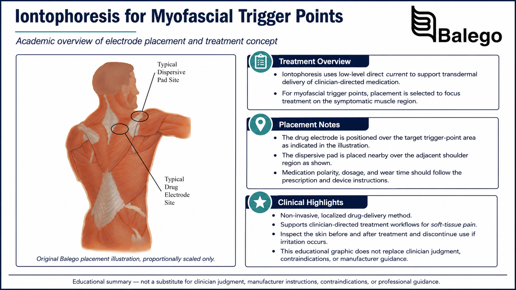
Myofascial Trigger-Point Iontophoresis: Quick Summary
| Clinical question | Practical answer |
|---|---|
| What is a trigger point? | A hypersensitive spot within a taut band that reproduces recognizable local or referred symptoms |
| Is every tender muscle spot a trigger point? | No, diagnosis should use several compatible findings |
| What is an active trigger point? | A point associated with the patient’s spontaneous or familiar pain |
| What is a latent trigger point? | A point that is painful primarily when examined and may not reproduce the presenting complaint |
| Where is the medication electrode placed? | Over the clinically relevant hypersensitive spot when the electrode can maintain full safe contact |
| Where is the return electrode placed? | Over intact nearby muscle tissue according to the electrode-system instructions |
| Is iontophoresis proven for every trigger-point location? | No, direct research is concentrated on upper-trapezius trigger points |
| Which medication should be used? | Only the exact formulation selected by the prescriber |
| Is iontophoresis equivalent to an injection? | No |
| Should exercise accompany treatment? | Yes, load management, movement, strengthening, and contributing-factor correction should remain central |
| When should treatment stop? | When skin irritation develops, the diagnosis becomes uncertain, or no measurable functional benefit occurs |
What Is Myofascial Pain Syndrome?
Myofascial pain syndrome is generally described as localized or regional muscle pain associated with one or more clinically relevant trigger points. Unlike widespread pain conditions, symptoms are often concentrated within a particular muscle or related group of muscles.
Potential features include:
- Deep aching pain
- A taut or rope-like muscle band
- A highly sensitive spot within that band
- Pain reproduced by compression
- Referred pain away from the point being pressed
- Reduced range of motion
- Pain with muscle contraction
- Perceived weakness
- Muscle guarding
- Headache or regional symptoms linked to a neck or shoulder muscle
- Symptoms that increase with sustained posture or repetitive load
The biological mechanisms remain under investigation. Current models include altered motor-endplate activity, sustained local contraction, reduced local circulation, nociceptor sensitization, and interaction with broader nervous-system and psychosocial factors. Myofascial pain should therefore be assessed within a biopsychosocial framework rather than attributed to a single isolated “knot.”
Active vs. Latent Trigger Points
| Feature | Active trigger point | Latent trigger point |
|---|---|---|
| Spontaneous symptoms | May reproduce the patient’s current pain | Usually not spontaneously painful |
| Pain with pressure | Yes | Yes |
| A familiar, referred pattern | Often present | May occur only during examination |
| Functional relevance | More likely to explain the presenting complaint | May represent a secondary finding |
| Treatment priority | Higher when examination findings are consistent | Treat only when clinically relevant |
| Diagnostic caution | Must still be differentiated from nerve, joint, tendon, or systemic pain | Should not be assumed to require treatment |
The distinction matters because the presence of multiple tender spots does not mean every spot should receive medication, needling, massage, or another procedure.
How Should a Trigger Point Be Diagnosed?
No single examination sign is sufficiently definitive by itself.
A 2023 diagnostic study found that the most commonly identified classical findings included the following:
- A taut band
- A hypersensitive spot within the band
- A possible palpable nodule
- Referred pain
The authors proposed that a more definite clinical diagnosis should require a hypersensitive spot within a taut band plus referred pain, a local twitch response, or multiple complementary signs of muscular dysfunction.
Practical diagnostic cluster
| Finding | Clinical value |
|---|---|
| Palpable taut band | Suggests localized altered muscle tension |
| Hypersensitive point within the band | Helps localize the suspected trigger point |
| Familiar local pain | Links the examination finding to the patient’s complaint |
| Referred pain | Strengthens the trigger-point interpretation |
| Restricted movement | Demonstrates functional effect |
| Pain with contraction | Supports involvement of the examined muscle |
| Local twitch response | May support the diagnosis but is not always present |
| Jump sign | Indicates marked tenderness but is nonspecific |
| Weakness or altered recruitment | May show neuromuscular dysfunction |
| Symptom change after targeted treatment | Can support—but does not independently prove—the diagnosis |
A taut band alone is not sufficient. A tender point outside a taut band may represent general muscle tenderness, another soft-tissue problem, or referred pain from another structure.
Common Contributing Factors
Myofascial symptoms may develop or persist because of several interacting factors.
| Category | Examples |
|---|---|
| Acute overload | Lifting injury, collision, sudden stretch, unfamiliar exercise |
| Repetitive load | Repeated reaching, gripping, typing, overhead work, running |
| Sustained posture | Prolonged sitting, static neck position, unsupported arms |
| Deconditioning | Low muscle endurance or insufficient load capacity |
| Altered movement | Compensation after injury, surgery, or joint restriction |
| Sleep and recovery | Insufficient sleep, poor recovery, high training volume |
| Psychological load | Stress, anxiety, fear, pain-related guarding |
| Ergonomics | Poor workstation setup, unsuitable tools, repeated awkward position |
| Regional impairment | Joint stiffness, reduced mobility, weakness, nerve sensitivity |
| Systemic contributors | Endocrine, metabolic, nutritional, inflammatory, or medication-related factors |
The goal is not merely to locate a painful point. The clinician must identify why the muscle is overloaded, guarded, or unable to recover.
What Does the Evidence Say About Iontophoresis?
Direct evidence is limited and should be interpreted narrowly.
2021 magnesium-sulfate study
A randomized study enrolled 60 participants, ages 19–24, with active trigger points in the dominant upper trapezius. The iontophoresis group received magnesium-sulfate iontophoresis twice weekly for four weeks, while the comparison group received direct current without medication.
The researchers reported improvements in pain, pressure-pain threshold, neck movement, and function in the medication group. The study involved one muscle region, a young population, one medication formulation, and relatively short follow-up. It does not establish that magnesium sulfate—or iontophoresis generally—is effective for every muscle or form of chronic myofascial pain.
2024 randomized trial
A later trial studied 75 young male physical-therapy students with mechanical neck pain and bilateral upper-trapezius trigger points. Participants received:
- Magnesium-sulfate iontophoresis plus conventional therapy
- High-power pain-threshold ultrasound plus conventional therapy
- Conventional therapy alone
Both modality groups improved more than the conventional-therapy group in several short-term outcomes, but iontophoresis was not superior to high-power ultrasound. The authors also identified short follow-up as a limitation.
Evidence limitations
| Limitation | Why it matters |
|---|---|
| Studies focused on upper trapezius | Results should not automatically be applied to lumbar, gluteal, calf, jaw, or forearm muscles |
| Participants were young adults | Findings may not generalize to older adults or complex chronic-pain populations |
| Magnesium sulfate was studied | Results do not establish effectiveness of corticosteroids, anesthetics, or other drugs |
| Treatment was combined with exercise or conventional therapy | Improvement cannot be attributed solely to iontophoresis |
| Follow-up was short | Long-term effectiveness and recurrence are uncertain |
| Trigger-point diagnosis varies | Study populations may not match every clinic’s diagnostic criteria |
| Direct comparison evidence is limited | Superiority over exercise, manual therapy, needling, or injection is not established |
Evidence-aware conclusion
Iontophoresis may be considered for short-term symptom modification in selected patients—particularly when an upper-trapezius trigger point and prescribed ionic medication resemble the studied circumstances.
Evaluate the Patient Before Treatment
Subjective history
Document:
| Question | Why it matters |
|---|---|
| Where is the primary pain? | Defines the regional complaint |
| Is the pain local or referred? | Helps test whether the suspected point reproduces the familiar pattern |
| How did symptoms begin? | Differentiates acute overload, trauma, and gradual-onset pain |
| What positions or activities aggravate symptoms? | Identifies the perpetuating load |
| Is pain constant or intermittent? | Helps assess irritability and differential diagnosis |
| Is numbness or tingling present? | Raises concern for nerve involvement |
| Is pain widespread? | May indicate a broader pain condition rather than isolated MPS |
| Are fatigue, sleep disturbance, or cognitive symptoms prominent? | May suggest fibromyalgia or systemic contributors |
| Is weakness true or pain-limited? | Helps distinguish neurologic and muscle causes |
| Is there unexplained weight loss, fever, or night pain? | Screens for medical red flags |
| What treatment has already been attempted? | Prevents indefinite repetition of ineffective care |
| What medications and allergies are present? | Required before iontophoresis |
| Is there an implanted electronic device? | May affect suitability for direct-current treatment |
| What function must improve? | Establishes meaningful outcomes |
Observation
Assess:
- Resting posture
- Protective movement
- Muscle asymmetry
- Swelling or discoloration
- Scarring
- Skin condition
- Breathing pattern
- Regional movement strategy
- Work or sport posture when relevant
Postural findings should not be treated as proof of causation. They are useful only when they reproduce symptoms or demonstrate a meaningful loading problem.
Palpation
Palpate systematically rather than searching only for the most painful point.
Document:
- Muscle examined
- Taut band
- Hypersensitive point within the band
- Familiar local pain
- Referred symptoms
- Local twitch response
- Tissue temperature or swelling
- Whether the finding is superficial enough for safe electrode placement
- Whether nearby nerves, vessels, organs, or bony prominences affect treatment selection
Repeated aggressive palpation can increase pain and sensitization. Use only the pressure necessary to establish a reproducible finding.
Movement and strength
Assess:
- Active range of motion
- Passive range when relevant
- Pain during contraction
- Pain during stretch
- Muscle endurance
- Load tolerance
- Functional movement
- Adjacent joint mobility
- Regional strength
- Neurologic status where indicated
A trigger point may be secondary to another impairment. For example, an upper-trapezius point may coexist with cervical radiculopathy, shoulder weakness, scapular-control deficits, or sustained occupational load.
Differential Diagnosis
Myofascial pain is a diagnosis of clinical reasoning—not simply palpation.
| Possible alternative | Findings that may distinguish it |
|---|---|
| Cervical or lumbar radiculopathy | Dermatomal symptoms, neurological weakness, altered reflexes, nerve-tension findings |
| Peripheral nerve entrapment | Numbness, tingling, sensory change, weakness in a named nerve distribution |
| Tendinopathy | Pain concentrated at a tendon and reproduced with tendon loading |
| Joint disorder | Joint-line pain, stiffness, swelling, mechanical restriction |
| Muscle strain | Acute onset, bruising, weakness, pain during contraction and stretch |
| Fibromyalgia | Widespread pain with fatigue, sleep disturbance, and broader sensory sensitivity |
| Inflammatory disease | Morning stiffness, joint swelling, systemic symptoms |
| Infection | Fever, redness, warmth, progressive swelling |
| Vascular disorder | Swelling, color change, abnormal pulses, exertional symptoms |
| Complex regional pain syndrome | Disproportionate pain, autonomic changes, temperature or color asymmetry |
| Referred visceral pain | Symptoms inconsistent with mechanical muscle loading |
| Medication-related myalgia | Temporal relationship to a medication |
| Stress fracture or tumor | Focal bone pain, night pain, progressive unexplained symptoms |
Imaging does not routinely confirm myofascial pain syndrome. It may be used when the history or examination suggests another diagnosis.
Red Flags and Referral Criteria
Obtain medical evaluation before iontophoresis when there is:
- Unexplained fever
- Significant redness or swelling
- Suspected infection
- Progressive neurologic weakness
- Loss of sensation
- New bowel or bladder dysfunction
- Severe unremitting night pain
- Unexplained weight loss
- Major trauma
- Suspected fracture or muscle rupture
- Vascular symptoms
- Widespread pain with systemic features
- A palpable enlarging mass
- Chest pain or shortness of breath
- A new severe headache with neurologic signs
- Symptoms inconsistent with a regional muscular presentation
- Uncertainty about the medication or its polarity
- A contraindicated implanted electronic device
- Damaged skin at the planned electrode site
When Iontophoresis May Fit
A clinician may consider iontophoresis when:
- A clinically relevant active trigger point is reasonably identified.
- The target lies in a superficial muscle accessible through intact skin.
- Symptoms are regional rather than unexplained and widespread.
- The medication has been selected by a prescriber.
- The medication is water-soluble and ionically appropriate.
- Polarity is verified.
- The electrode can maintain full safe contact.
- The patient prefers or requires a non-needle option.
- Short-term symptom relief would help participation in exercise or functional retraining.
- A defined reassessment point has been established.
When another approach may be preferable
| Finding | Better next step |
|---|---|
| Prominent numbness or tingling | Complete neurologic evaluation |
| Deep muscle beneath substantial tissue | Consider whether surface iontophoresis can reasonably target the region |
| Widespread pain and fatigue | Evaluate for fibromyalgia or systemic contributors |
| Acute muscle tear | Protect, assess, and rehabilitate the injury |
| Uncertain tender-point diagnosis | Reassess before applying medication |
| Multiple perpetuating ergonomic factors | Correct loading and work setup |
| Severe fear and activity avoidance | Use graded exposure and pain education |
| No response to a defined trial | Stop repeating the modality and reconsider the plan |
| Skin reaction or poor electrode contact | Select another format or treatment |
| Need for direct mechanical intervention | Consider qualified manual therapy, needling, or medical referral |
Iontophoresis Protocol Framework
Step 1: Confirm the medication order
Verify:
- Medication name
- Exact chemical formulation
- Concentration
- Prescribed volume
- Ionic charge
- Required electrode polarity
- Electrical dose
- Maximum current
- Treatment frequency
- Planned treatment series
- Medication allergies
- Treatment location
Do not assume that a medication is appropriate because it was used in a study of a different muscle or population.
Magnesium sulfate has been studied in upper-trapezius trigger points, but that research does not create a universal magnesium-sulfate protocol. Corticosteroid iontophoresis should likewise not be used without an exact prescriber order.
Step 2: Establish baseline outcomes
Choose measures that match the affected region.
| Outcome | Possible measure |
|---|---|
| Pain intensity | 0–10 numerical rating |
| Pressure sensitivity | Pressure algometer when available |
| Familiar referred pain | Standardized palpation response |
| Range of motion | Region-specific goniometric measurement |
| Muscle endurance | Timed or repetition-based test |
| Strength | Handheld dynamometry or standardized resistance |
| Function | Patient-Specific Functional Scale |
| Neck function | Neck Disability Index |
| Upper-extremity function | QuickDASH |
| Low-back function | Oswestry Disability Index |
| Activity tolerance | Time or repetitions during the provoking task |
| Sleep effect | Patient-reported sleep interference |
| Skin status | Inspection before every treatment |
Pressure-pain threshold may improve even when function does not. Use more than one outcome.
Step 3: Identify the treatment target
Mark the point before preparing the electrode.
The target should demonstrate:
- A hypersensitive spot
- Location within a taut band
- Reproduction of familiar symptoms
- A clinically relevant local or referred pattern
- Relationship to the functional complaint
- A safe surface for electrode contact
Avoid treating a point merely because it is painful when pressed.
Step 4: Choose an electrode format
Consider:
- Size of the symptomatic region
- Muscle contour
- Medication volume
- Required dose
- Skin sensitivity
- Hair
- Patient position
- Need for current adjustment
- Proximity to nerves or bony prominences
- Whether the patient can remain still
- Whether a wearable patch can remain secure
A small flexible electrode may suit a localized upper-trapezius or forearm target. A broader or butterfly shape may fit larger curved muscle regions.
Step 5: Prepare the skin according to the product
Do not apply one skin-preparation instruction to every iontophoresis system.
Some controller-based electrode kits include alcohol preparation pads. Wearable systems may direct users to clean with soap and water instead.
General principles include:
- Use intact skin.
- Remove lotion, oil, and heavy contamination.
- Dry the site completely.
- Clip excessive hair if necessary.
- Do not shave immediately before treatment.
- Remove jewelry near the circuit.
- Avoid wounds, rashes, recent scars, and irritated skin.
- Inspect both medication and return-electrode sites.
Use Iontophoresis Electrode Directions for Use for the selected controller-electrode workflow.
Step 6: Position the medication electrode
Place the medication electrode.
- Directly over the marked clinically relevant point
- With the conductive reservoir centered on the target
- With the full electrode surface contacting the skin
- Away from damaged skin
- Away from a prominent superficial nerve when possible
- Away from excessive pressure from clothing, straps, or body weight
- Without wrinkles or lifted edges
- According to the verified medication polarity
The deepest tender point cannot always be reached effectively through surface iontophoresis. Electrode placement should be based on anatomy and safe contact—not on pressing a reservoir as deeply as possible into the tissue.
Step 7: Position the return electrode
Place the return electrode:
- On intact nearby muscle tissue
- According to the selected electrode’s spacing requirements
- Away from prominent bone
- Where the full surface remains adhered
- Without crossing the chest
- Without creating a current path through a contraindicated region
- Where clothing or body weight will not compress it
The return electrode may be placed proximal or distal depending on the region and product instructions. There is no universal “nearest muscle mass” position for every trigger point.
Step 8: Support the patient and minimize movement
During controller-based delivery:
- Support the treated region comfortably.
- Keep the target muscle relaxed when practical.
- Provide adequate lead-wire slack.
- Do not allow cables to pull on the electrodes.
- Avoid repeated movement that lifts the pads.
- Do not place the patient’s body weight on either electrode.
- Keep clothing from compressing or rubbing the setup.
Exercise and movement can be performed before or after treatment. Repeated movement during active delivery should occur only when expressly permitted by the device and clinician protocol.
Step 9: Deliver the prescribed treatment
Controller-based iontophoresis
The ActivaDose II Iontophoresis Controller allows the clinician to select an ordered dose from 0–80 mA·min, adjust current up to 4 mA, and monitor dose, current, time, resistance, and electrode alerts.
General workflow:
- Connect the electrodes according to verified polarity.
- Enter the prescribed electrical dose.
- Select a tolerable current within the electrode limits.
- Begin using automatic ramp-up.
- Monitor the patient and device indicators.
- Reduce current if excessive stinging develops.
- Stop for burning, sharp pain, loss of contact, or repeated electrode rejection.
- Allow the controller to ramp down at completion.
A 40 mA·min dose should not be called the universal trigger point dose. The current live page’s reference to “40 mA-min” appears as a keyword rather than a properly sourced prescription.
Wearable iontophoresis patch
With a self-contained patch:
- Confirm the prescribed patch model and dose capacity.
- Identify the correct medication chamber.
- Apply only the directed medication volume.
- Complete the model-specific activation procedure.
- Center the medication chamber over the selected point.
- Confirm that the patch does not restrict movement or compress a nerve.
- Record application and planned removal time.
- Provide written skin and removal instructions.
- Do not extend wear beyond the device instructions.
- Discard after one use.
Step 10: Inspect the skin
After treatment:
- Reduce controller current to zero when applicable.
- Remove the electrode or patch according to its instructions.
- Inspect both sites.
- Record redness, itching, burning, blistering, or adhesive reaction.
- Identify whether the reaction occurred under the medication or return electrode.
- Do not immediately treat irritated skin.
- Report suspected medication reactions.
Iontophoresis can cause irritation or burns, especially when contact is uneven, current density is high, the skin is damaged, or the patient cannot accurately report sensation.
Placement Considerations by Muscle Region
This table describes fit and safety considerations—not medication prescriptions.
| Muscle region | Possible electrode consideration | Important caution |
|---|---|---|
| Upper trapezius | Flexible electrode over the reproducible point; return electrode on nearby shoulder or upper-back muscle | Avoid the anterior or lateral neck and do not create a current path across the head or chest |
| Levator scapulae | A small or form-fitting electrode may fit the superior medial scapular region | Differentiate cervical referral and dorsal scapular nerve symptoms |
| Infraspinatus | Electrode over the posterior scapular muscle belly | Confirm symptoms are not primarily glenohumeral or cervical |
| Forearm flexors or extensors | Small electrode over a localized superficial point | Screen for tendon and peripheral-nerve disorders |
| Lumbar paraspinals | A larger electrode may be required | Rule out radiculopathy, fracture, visceral referral, and red flags |
| Quadratus lumborum | The target can be deep and anatomically variable | Avoid assuming surface delivery reaches a deep point |
| Gluteal muscles | A larger flexible or butterfly electrode may improve contact | Screen for lumbar, hip-joint, and sciatic-nerve causes |
| Piriformis region | The surface target is deep beneath gluteal tissue | Confirm diagnosis and recognize limited surface-delivery specificity |
| Calf | An electrode may fit the muscle belly | Rule out deep-vein thrombosis, vascular disease, strain, or nerve involvement |
| Masseter or temporalis | Small target near sensitive facial anatomy | Follow facial and orbital contraindications; specialist guidance is appropriate |
Direct iontophoresis evidence should not be inferred merely because a trigger-point chart names a muscle.
Comparing Balego Iontophoresis Options
The full specifications remain on the linked product pages. This comparison focuses on trigger-point workflow.
| Balego option | Format | Potential trigger-point advantage | Important limitation |
|---|---|---|---|
| ActivaDose II Controller | Reusable external controller | Direct current and dose control: treatment can be monitored in the clinic. | Requires compatible disposable electrodes and limited movement |
| Trivarion Delivery Kits | Flexible controller-based electrode | Form-fitting construction helps on curved shoulder, elbow, and other uneven sites | Electrode size must match the target and medication volume |
| Ionto4 Electrode Kits | Controller-based square or butterfly system | Small, medium, large, and butterfly options support different muscle regions | A flat square may not fit every contour |
| Buffered Iontophoresis Electrode Kits | Controller-based Ag/AgCl system | Multiple sizes and a buffered return-electrode design | Requires external controller and lead wires |
| IontoPATCH | Self-contained wearable patch | No external controller; 80- and 120-mA·min options | The patch footprint may be too large for a small point, and medication choice must still be prescribed |
| ACTIVApatch | Self-contained no-saline patch | Treatment options from about 1.25 to 12 hours with no external wires | Fit, pressure, and motion must be assessed for each muscle region |
Trivarion is available in small, medium, butterfly, and large formats and is designed to conform to curved or uneven treatment areas. Ionto4 and Balego’s buffered electrode family provides several square and butterfly sizes.
Best format by clinical priority
| Priority | Format to consider |
|---|---|
| First application with close observation | ActivaDose II with a compatible electrode |
| Small, localized point | Small Trivarion or Ionto4 electrode |
| Curved shoulder or scapular region | Trivarion or butterfly electrode |
| Exact current adjustment | ActivaDose II |
| Patient has uncertain skin tolerance | Controller-based treatment under direct observation |
| Short wearable treatment | ACTIVApatch IntelliDose 2.5 or another prescribed short-wear model |
| Standard wearable 80 mA·min dose | IontoPatch or ACTIVApatch model selected by schedule and fit |
| Higher 120 mA·min patch capacity | IontoPatch Extra Strength only when specifically ordered |
| Multiple unrelated trigger points | Treat selectively rather than placing patches on every tender spot |
IontoPatch models provide 80- or 120-mA·min capacity with average wear periods of approximately 4, 8, or 14 hours. ACTIVApatch models provide 40- or 80-mA·min options over approximately 1.25 to 12 hours. Medication is not included with either product family.
Iontophoresis Compared with Other Treatments
| Treatment | Primary role | Potential advantage | Important limitation |
|---|---|---|---|
| Education and load modification | Reduces perpetuating stress | Addresses contributing factors | Requires behavior change |
| Progressive exercise | Restores strength, endurance, and movement capacity | Central to long-term function | Must be individually progressed |
| Manual pressure or soft-tissue treatment | Short-term symptom and mobility modification | Noninvasive and easy to combine with exercise | Effects may be temporary |
| Stretching | Addresses relevant muscle restriction | Accessible home intervention | Aggressive stretching can aggravate symptoms |
| Dry needling | Direct needling of a clinically selected point | May provide short-term pain relief in some conditions | Invasive, scope-dependent, and not universally superior |
| Acupuncture | Needle-based neuromodulatory intervention | May reduce pain in selected cases | Protocols and evidence vary |
| Trigger-point injection | Delivers local anesthetic or another medication through a needle | Direct placement into the selected tissue | Invasive and associated with site-specific procedural risks |
| Iontophoresis | Non-needle transdermal medication delivery | Controlled current and no needle puncture | Variable delivery, skin reactions, and limited condition-specific evidence |
| Therapeutic ultrasound | Physical agent sometimes used for short-term symptoms | Noninvasive | Evidence and treatment parameters vary |
| Laser or shockwave | Selected physical-agent options | Noninvasive | Availability, protocols, and evidence differ |
| Heat or cold | Temporary symptom management | Accessible and inexpensive | Does not address the primary loading problem |
| Medication | Symptom management | May reduce pain enough to permit activity | Depends on medical suitability and side effects |
Iontophoresis avoids a needle puncture, but that does not make it equivalent to a trigger-point injection. Injections place medication within or near the targeted tissue, whereas iontophoresis relies on movement through the skin and superficial tissues.
Rehabilitation That Should Accompany Treatment
Iontophoresis should support—not replace—active rehabilitation.
Phase 1: Reduce irritability
Possible strategies include:
- Modify the provoking task.
- Break up sustained postures.
- Use a comfortable active range of motion.
- Begin low-load isometric exercise.
- Improve breathing and reduce unnecessary guarding.
- Address sleep and recovery.
- Use short-duration manual treatment when helpful.
- Educate the patient that temporary tenderness does not necessarily indicate tissue damage.
Phase 2: Restore movement and capacity
Progress toward:
- Full comfortable range of motion
- Concentric and eccentric strengthening
- Regional endurance
- Scapular, trunk, or hip control when relevant
- Grip or lifting endurance
- Postural variation
- Gradual return to work or sport load
- Repeated exposure to previously provocative movements
Our resistance bands and tubing can support clinician-directed progressive strengthening.
Phase 3: Address the perpetuating factor
Examples include:
- Workstation modification
- Tool-handle changes
- Improved lifting strategy
- Training-volume adjustment
- Rest and recovery scheduling
- Shoulder or trunk strengthening
- Running-mechanics changes
- Graded return to throwing or overhead work
- Stress-management or relaxation strategies
- Treatment of an adjacent joint or nerve disorder
A successful plan should improve what the patient can do—not merely reduce tenderness when the point is pressed.
Reassessment and Stopping Rules
Recommended outcome measures
| Outcome | Baseline | Reassessment |
|---|---|---|
| Pain | 0–10 rating under defined conditions | Repeat under the same conditions |
| Pressure-pain threshold | Algometer reading when available | Same point and technique |
| Referred pain | Standardized palpation response | Compare intensity and distribution |
| Range of motion | Region-specific measure | Repeat same movement |
| Muscle endurance | Time or repetitions | Same position and load |
| Function | PSFS, QuickDASH, NDI, ODI, or other regional scale | Repeat the same instrument |
| Activity tolerance | Time, repetitions, or load | Repeat the same task |
| Sleep interference | Patient-reported frequency | Compare over time |
| Medication tolerance | Local or systemic response | Review after every treatment |
| Skin response | Visual inspection | Inspect both sites every session |
Continue only when:
- Skin remains suitable.
- The medication remains medically appropriate.
- Pain or function is improving.
- The patient is progressing with movement and exercise.
- The target remains clinically relevant.
- Iontophoresis is serving a defined functional purpose.
Modify or discontinue when:
- Burning, blistering, or persistent irritation occurs.
- The electrode repeatedly loses contact.
- Symptoms become more widespread.
- Numbness or weakness develops.
- The suspected point no longer reproduces the familiar complaint.
- No measurable functional change occurs.
- Relief is brief and does not improve activity tolerance.
- The modality delays a needed diagnostic evaluation.
- Passive treatment is replacing rehabilitation.
- The patient requires repeated treatment of an expanding number of tender points.
Suggested Documentation Template
Evaluation
- Primary region:
- Muscle examined:
- Symptom duration:
- Familiar pain description:
- Local or referred pattern:
- Aggravating activities:
- Taut band present:
- Hypersensitive spot within band:
- Palpable nodule:
- Familiar pain reproduced:
- Referred symptoms reproduced:
- Local twitch response:
- Pain with contraction:
- Restricted movement:
- Strength or endurance deficit:
- Neurologic screen:
- Differential diagnoses:
- Skin condition:
- Functional baseline:
- Prior care:
Iontophoresis order
- Medication:
- Exact formulation:
- Concentration:
- Prescriber:
- Volume:
- Ionic charge:
- Polarity:
- Electrical dose:
- Maximum current:
- Frequency:
- Planned treatment trial:
- Device or patch:
- Active-electrode size:
- Active-electrode location:
- Return-electrode location:
Treatment response
- Current achieved:
- Treatment duration:
- Device alerts:
- Sensation during treatment:
- Skin before treatment:
- Skin after treatment:
- Pain change:
- Pressure-pain threshold change:
- Movement change:
- Functional change:
- Exercise progression:
- Continue, modify, discontinue, or refer:
Frequently Asked Questions
Can iontophoresis remove a muscle knot?
No. Iontophoresis assists transdermal delivery of an ionic medication. It does not physically break apart a knot or guarantee elimination of a trigger point.
Is iontophoresis proven for myofascial pain syndrome?
Evidence is limited. Small trials involving magnesium-sulfate iontophoresis have reported short-term improvements in young adults with upper-trapezius trigger points and neck pain. The evidence should not be generalized to every muscle or patient.
Which medication should be used?
Only an exact medication formulation selected by a prescriber. Magnesium sulfate has been studied in upper-trapezius research, but it is not a universal trigger-point medication.
Can dexamethasone be used?
Only when prescribed and when the exact formulation, concentration, polarity, volume, and dose have been confirmed. Evidence from another diagnosis should not be treated as proof for myofascial pain syndrome.
Where is the medication electrode placed?
It is generally centered over the clinically relevant hypersensitive spot within the taut band, provided the electrode can maintain full safe contact.
Where is the return electrode placed?
On intact nearby muscle tissue according to the selected electrode and controller instructions.
Is a 40 mA·min dose standard?
No universal trigger-point dose has been established. Follow the medical order and the limits of the selected electrode and controller.
How many treatments are needed?
No universal number has been established. Published magnesium-sulfate studies used twice-weekly treatment for four weeks, but that schedule should not be converted into a general prescription.
Can multiple trigger points be treated at once?
Only when medically and technically appropriate. Treating every tender point can increase medication exposure, skin burden, treatment complexity, and the risk of missing a broader diagnosis.
Should the muscle move during treatment?
During controller-based treatment, the region is generally supported to preserve electrode contact. Exercise is usually completed before or after active delivery unless the device and protocol specifically permit movement.
Is iontophoresis better than dry needling?
Current evidence does not establish universal superiority. The techniques differ substantially, and selection depends on diagnosis, patient preference, medication order, clinician training, and treatment goals.
Is iontophoresis better than a trigger-point injection?
No direct evidence establishes equivalence. Iontophoresis avoids a needle but provides variable transdermal delivery. Injection directly places medication in or near the selected tissue and carries different risks.
Can IontoPatch be placed over a trigger point?
Possibly, when the patch fits the anatomy, the medication is prescribed, and the reservoir can remain centered without excessive pressure or motion.
Is a wearable patch better than a controller?
Not universally. A controller offers direct current adjustment and observation. A wearable patch supports mobility and extended delivery but provides less real-time control.
When should the patient be referred?
Refer when symptoms are unexplained, widespread, progressive, neurologic, systemic, traumatic, associated with red flags, or unresponsive to an appropriate rehabilitation plan.
Related Resources
- Shop iontophoresis patches, electrodes, and dose controllers
- Iontophoresis Treatment Protocol Library
- Iontophoresis Electrode Directions for Use
- Iontophoresis FAQ, Medication, Dose, and Polarity Guides
- ActivaDose II Iontophoresis Controller
- Trivarion Form-Fitting Iontophoresis Electrodes
- Ionto4 Iontophoresis Electrode Kits
- Buffered Iontophoresis Electrode Kits
- IontoPatch Wearable Iontophoresis Patches
- ACTIVApatch Wearable Iontophoresis Patches
- Resistance Bands and Tubing
- Hot and Cold Therapy Products
This protocol is intended for professional education. Medication iontophoresis should be performed only under appropriate medical direction and according to the medication labeling, controller instructions, electrode or patch instructions, facility policy, and applicable scope-of-practice


