



Patellar tendinopathy is a load-related condition involving pain and reduced capacity in the tendon connecting the patella to the tibia. It is often called patellar tendonitis or jumper’s knee, particularly when symptoms develop during sports involving repeated acceleration, jumping, and landing.
The term "tendinopathy" is generally more accurate than "tendonitis." Persistent tendon pain is not explained solely by acute inflammation; tendon structure, load tolerance, pain sensitivity, muscle capacity, and training demands may all contribute. Diagnosis is primarily clinical, with imaging used selectively when the presentation is uncertain or another injury is suspected.
Iontophoresis may be considered when a qualified healthcare professional identifies a localized, superficial treatment target and a prescriber orders a compatible ionized medication. Its potential role is short-term symptom modification. It should not replace education, load management, progressive quadriceps and tendon loading, or a structured return to activity.
Clinical notice: Medication, concentration, polarity, dose, current, electrode selection, placement and treatment frequency require an appropriate order, professional judgment and compliance with the medication, controller, patch and electrode instructions for use.
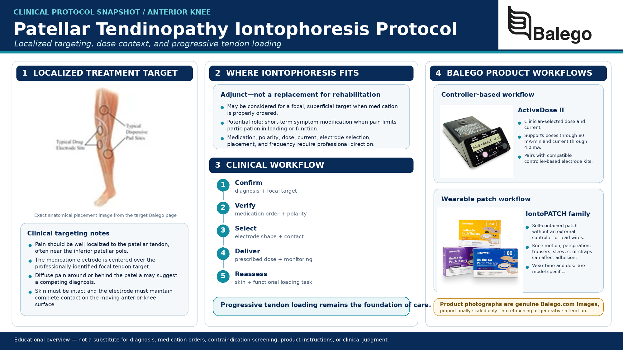
Patellar Tendinopathy Iontophoresis at a Glance
| Clinical question | Practical answer |
|---|---|
| Is all pain below the kneecap patellar tendinopathy? | No. Patellofemoral pain, fat pad irritation, tendon tears, bursitis, apophyseal conditions, and other injuries may produce symptoms in the same region. |
| What presentation best fits patellar tendinopathy? | Load-related pain that is well localized to the patellar tendon and reproduced by tendon-loading activity. |
| What is the main rehabilitation treatment? | Education, activity load management, and progressive tendon-loading exercise. |
| Where may iontophoresis fit? | As a prescribed adjunct when focal symptoms prevent participation in loading or functional activity, iontophoresis may be indicated. |
| Where is the drug electrode placed? | The drug electrode is placed over the professionally identified tendon target, which is commonly located near the inferior patellar pole or another focal symptomatic region. |
| Is 40 mA-min always the correct dose? | No. The principal patellar-tendinopathy trial used 80 mA-min, but current dosing must follow the prescription and product instructions. |
| Should activity stop completely? | Usually not. Provocative load is modified while tolerable strength and conditioning activities are maintained. |
| What determines readiness to return? | Tendon-loading capacity, strength, jumping or running tolerance, sport demands, and next-day symptom response—not just rest pain—determine readiness to return. |
What Does Patellar Tendinopathy Usually Feel Like?
A typical presentation includes:
- Pain localized to the patellar tendon
- Symptoms commonly near the inferior pole of the patella
- Pain during jumping, landing, accelerating or changing direction
- Pain during squats, stairs or loaded knee extension
- Symptoms that increase as tendon-loading demand increases
- Local tenderness
- Reduced tolerance for training or physically demanding work
- Morning or post-activity stiffness in some patients
Patellar tendinopathy is especially associated with sports that repeatedly expose the knee extensor mechanism to high loading rates, but it may also affect recreationally active people and workers performing repeated squatting, climbing, or lifting.
Pain at rest or generalized swelling or broad pain around the patella is less characteristic and should prompt careful consideration of competing diagnoses.
Patellar Tendinopathy vs. Other Causes of Anterior Knee Pain
The medication electrode should not be positioned until the clinician has identified a credible treatment target.
| Possible condition | Distinguishing considerations |
|---|---|
| Patellofemoral pain | Pain is often diffuse around or behind the patella and may be reproduced by squatting, stairs or prolonged sitting rather than focal tendon palpation |
| Patellar tendon partial or complete tear | Sudden injury, popping, bruising, weakness, tendon defect or loss of active knee extension |
| Quadriceps tendinopathy | Symptoms are concentrated above the patella rather than below it |
| Infrapatellar fat-pad irritation | Pain may be located on either side of the tendon and aggravated by knee extension or local compression |
| Prepatellar or infrapatellar bursitis | More superficial swelling and pressure-related tenderness may predominate |
| Osgood-Schlatter disease | Skeletally immature patient with symptoms at the tibial tubercle |
| Sinding-Larsen-Johansson syndrome | Skeletally immature patient with symptoms at the inferior patellar pole |
| Patellar stress injury | Focal bone pain, impact intolerance or symptoms inconsistent with isolated tendon loading |
| Meniscal or intra-articular disorder | Joint-line pain, effusion, catching, locking or rotational mechanism |
| Patellar instability | Apprehension, giving way or a history of patellar displacement |
| Inflammatory or infectious disorder | Marked warmth, redness, systemic symptoms or multi-joint involvement |
| Referred pain | Hip, lumbar or neurological findings influence the symptoms |
Patellar tendinopathy and patellofemoral pain may both hurt during squatting or stairs, but patellar tendinopathy is more strongly characterized by localized tendon pain associated with tendon-loading activity. A diagnosis should not be based on the phrase “pain below my kneecap” alone.
Red Flags and Findings Requiring Referral
Do not proceed with a routine tendinopathy protocol when the patient has:
- A sudden pop or tearing sensation
- Inability to actively extend the knee
- Inability to perform a straight-leg raise
- A palpable tendon defect
- Rapid bruising or substantial swelling after injury
- Acute traumatic deformity
- A large or rapidly developing joint effusion
- Fever, redness or a hot swollen joint
- Progressive neurological loss
- Unexplained night pain, systemic illness or weight loss
- Inability to bear weight after significant trauma
- Symptoms in a skeletally immature patient that have not been appropriately evaluated
Suspected disruption of the knee extensor mechanism requires prompt medical evaluation rather than iontophoresis or routine loading exercise.
Patellar Tendinopathy Examination Checklist
1. Clarify the symptom history
Document:
- Exact pain location
- Gradual or sudden onset
- Duration and progression
- Training and competition schedule
- Changes in jumping, sprinting or running volume
- Recent increases in intensity, surface or frequency
- Strength-training changes
- Work demands
- Pain during activity
- Pain after activity and during the following day
- Morning stiffness
- Previous knee injuries
- Prior rehabilitation or injection history
- Current medications and allergies
- Relevant inflammatory, metabolic or systemic conditions
- Patient goals and performance requirements
Avoid assuming that pronation, patellar alignment, or one anatomical feature caused the condition. These findings may be considered within the full examination, but they do not independently establish the diagnosis or treatment plan.
2. Map the pain precisely
Ask the patient to identify the pain with one finger.
Determine where symptoms are located:
- At the inferior patellar pole
- Within the tendon midsubstance
- At the tibial insertion
- Beside rather than within the tendon
- Behind or around the patella
- At the tibial tubercle
- At the superior patellar pole
The smaller and more reproducible the symptomatic tendon region, the more plausible it is as a localized iontophoresis target.
3. Inspect and palpate
Compare sides for:
- Local thickening
- Swelling
- Bruising
- Skin irritation
- Previous surgical scars
- Tendon continuity
- Tenderness at the inferior patellar pole
- Midsubstance or distal-tendon tenderness
- Adjacent fat-pad or bursal tenderness
- Sensation and skin suitability at proposed electrode sites
Palpation supports the diagnosis but should not be interpreted without load-related symptom reproduction.
4. Reproduce symptoms with tendon loading
Depending on irritability and safety, useful loading tasks may include the following:
- Squat
- Single-leg squat
- Decline squat
- Step-down
- Resisted knee extension
- Jump
- Landing
- Running or acceleration
- Sport-specific movement
Record:
- Exact pain location
- Pain intensity
- Movement strategy
- Load or depth at symptom onset
- Performance compared with the other side
- Symptom response after the test
A single-leg decline squat is commonly used to assess load-related patellar-tendon pain, but no single test should replace the complete clinical examination.
5. Measure strength and capacity
Assess as appropriate:
- Quadriceps force
- Knee-extension tolerance
- Squat strength
- Single-leg squat control
- Hip strength
- Calf capacity
- Repeated jump or hop capacity
- Landing control
- Rate and quality of force production
- Relevant work or sport tasks
Strength symmetry alone is insufficient. Tendon rehabilitation must also prepare the patient for the speed, repetition, and loading rate of the required activity.
6. Assess mobility when clinically relevant
Consider:
- Knee flexion and extension
- Ankle dorsiflexion
- Hip motion
- Quadriceps flexibility
- Hamstring flexibility
- Calf flexibility
Mobility findings should guide treatment only when they meaningfully relate to the patient’s symptoms, movement demands, or rehabilitation progression.
7. Establish validated and functional baselines
Useful measures include:
- Victorian Institute of Sport Assessment–Patella, or VISA-P
- Patient-Specific Functional Scale
- Pain during a standardized loading task
- Single-leg decline-squat response
- Knee-extension strength
- Squat load and depth
- Jump height or repeated-jump tolerance
- Running volume
- Training participation
- Work tolerance
VISA-P is commonly used to quantify patellar-tendinopathy symptoms and functional limitation, while repeated functional loading tests help connect the score to the patient’s actual activity demands.
Is Imaging Necessary?
Patellar tendinopathy is usually diagnosed clinically. Ultrasound or MRI may support the examination when:
- The diagnosis remains uncertain
- A partial tear is suspected
- Symptoms followed a significant acute injury
- The clinical pattern is atypical
- Surgery or another procedure is being considered
- The patient has failed to progress despite a well-structured program
- Another tendon, bone or intra-articular disorder is suspected
Imaging abnormalities do not automatically identify the source of pain, and changes in tendon appearance do not always parallel clinical recovery. Recent research also indicates that the prognostic value of conventional ultrasound and MRI findings during exercise-based rehabilitation remains uncertain.
Treatment should therefore target the patient’s symptoms and functional capacity rather than attempting to “normalize” an image.
What Does the Evidence Say About Iontophoresis?
The most directly relevant trial was a blinded randomized clinical study involving 31 participants with patellar tendinopathy.
Participants were assigned to:
- Wireless dexamethasone iontophoresis
- Wired dexamethasone iontophoresis
- Sham treatment
The two active groups received six treatments using 2 mL of 4% dexamethasone sodium phosphate at an 80 mA-min dose. All three groups improved during the study. The active wired and wireless treatments produced greater pain reduction during a functional task, while broader outcome differences were limited. The authors concluded that the wireless patch performed similarly to the wired controller.
How to interpret that trial
| Finding | Clinical interpretation |
|---|---|
| Active treatment reduced functional-task pain more than sham | Iontophoresis may provide short-term symptom modification in selected cases |
| All groups improved | Improvement cannot be attributed solely to active medication delivery |
| Wired and wireless treatment performed similarly | Either workflow may be reasonable when it matches the prescription and treatment site |
| Six 80 mA-min treatments were used | This describes the research protocol, not a universal prescription |
| The study included only 31 participants | Findings should not be overstated or generalized to every anterior-knee condition |
| No progressive loading program was included | The trial does not show that iontophoresis is superior to comprehensive rehabilitation |
The study isolated iontophoresis rather than combining it with loading exercise. Modern patellar-tendinopathy management instead places education and progressive tendon loading at the center of care.
Where Iontophoresis May Fit
Iontophoresis may be considered when:
- The diagnosis is reasonably clear
- Symptoms are focal and superficial
- The skin is intact
- A medication has been appropriately ordered
- Pain limits participation in strengthening or functional activity
- The electrode can maintain complete contact on the anterior knee
- Knee motion and clothing pressure can be controlled
- A measurable treatment objective has been established
- The plan includes progressive loading and reassessment
It is less likely to be useful when:
- Pain is diffuse around or behind the patella
- The treatment target cannot be clearly localized
- A tendon tear is suspected
- The knee is substantially swollen
- Skin condition or sensation makes treatment unsafe
- The patient expects passive treatment to replace rehabilitation
- Previous treatments did not change symptoms or function
- The patch cannot be protected from knee sleeves, straps, braces or repeated flexion
Iontophoresis Is Not the Same as a Tendon Injection
Iontophoresis should not be described simply as a needle-free equivalent to injection.
| Feature | Iontophoresis | Injection |
|---|---|---|
| Skin puncture | No | Yes |
| Delivery route | Medication transport through intact skin using direct current | Medication introduced with a needle into or around a selected structure |
| Placement precision | Influenced by electrode position, skin, current and tissue properties | Influenced by needle position and whether imaging guidance is used |
| Evidence in patellar tendinopathy | One small randomized iontophoresis trial | Evidence varies by the injected substance and procedure |
| Main risks | Skin irritation, burns, medication reaction and electrical discomfort | Infection, bleeding, tissue injury and medication-related effects |
| Proven equivalent? | No | Direct equivalence has not been established |
Medication selection and procedural decisions should be made on their own clinical rationale.
Step-by-Step Patellar Tendinopathy Iontophoresis Protocol
Step 1: Confirm the treatment target
Document:
- Working diagnosis
- Exact symptomatic tendon region
- Findings supporting patellar tendinopathy
- Competing diagnoses considered
- Baseline functional test
- Reason iontophoresis is being added
Do not use “anterior knee pain” as the sole treatment indication.
Step 2: Define a functional objective
Examples include:
- Improve tolerance for introductory quadriceps loading
- Reduce pain during a squat
- Improve tolerance for stair climbing
- Improve participation in practice modification
- Reduce a symptom barrier during a return-to-running program
“Reduce inflammation” is not a complete functional goal.
Step 3: Verify the medication order
Confirm:
- Medication
- Formulation and concentration
- Medication polarity
- Prescribed dose
- Treatment frequency
- Allergies
- Patient-specific precautions
- Electrode or patch compatibility
Medication is not included with Balego’s iontophoresis controllers, electrodes, or patches.
For general explanations of direct current, medication polarity, and mA-min dosing, use our Iontophoresis Frequently Asked Questions rather than repeating that information on this condition-specific page.
Step 4: Inspect the skin
Inspect both electrode sites for:
- Cuts or abrasions
- Rash
- Folliculitis
- Recent shaving
- Scar irritation
- Altered sensation
- Bruising
- Significant swelling
- Previous electrode reactions
- Tape, brace or sleeve irritation
Do not apply an electrode over broken, infected, or significantly irritated skin.
Step 5: Select the electrode shape and size
The patellar tendon is a relatively narrow structure over a moving anterior-knee surface.
Choose an electrode that:
- Covers the focal target
- Holds the prescribed solution volume
- Maintains full adhesive contact
- Does not fold over the inferior patellar edge
- Does not extend into a deep knee crease
- Does not cover unnecessary skin
- Will not be compressed by clothing or a brace
A flexible or butterfly-shaped electrode may conform better than a square electrode, which would bridge the curved inferior-patellar region.
Step 6: Prepare the skin and electrodes.
Follow the exact product instructions.
General principles include:
- Remove lotion, oil and perspiration
- Clip excessive hair if needed
- Avoid shaving immediately before treatment
- Prepare the active electrode off the patient
- Hydrate the complete drug matrix
- Avoid dry areas
- Do not overfill or underfill
- Verify polarity before connecting the leads
Our Iontophoresis Electrode Directions for Use contains the complete filling, connection, polarity, dose limit, and safety instructions. Balego’s electrode directions specify single-use treatment kits, off-patient filling, and product-dependent limits for negative- and positive-polarity delivery.
Step 7: Position the active medication electrode
Place the medication reservoir over the clinically identified focal tendon target.
Common targets may include:
- The proximal tendon near the inferior patellar pole
- A focal midsubstance region
- A less common distal-tendon target near the tibial attachment
Placement principles:
- Center the drug matrix over the symptomatic tendon region
- Maintain complete skin contact
- Avoid wrinkling over the patellar edge
- Avoid excessive pressure directly over bone
- Keep the electrode away from irritated skin
- Do not assume every patient should receive identical inferior-pole placement
Step 8: Position the return electrode
With a controller-based system, place the return electrode on an appropriate area of intact soft tissue according to the product instructions.
A proximal thigh or other suitable ipsilateral soft-tissue location may be considered when it provides the following:
- Full electrode contact
- Adequate separation
- No clothing compression
- No interference from a knee sleeve
- No pressure from the treatment table
- Minimal lead-wire tension
Do not bind or tightly tape either electrode.
Step 9: Check knee position and contact
Support the leg in a comfortable, stable position. Before treatment:
- Gently check a small amount of knee motion
- Confirm the active electrode remains flat
- Reposition if an edge lifts
- Ensure the patient will not repeatedly flex and extend the knee
- Ensure clothing does not press over either electrode
- Keep lead wires free from tension
Excessive movement can disrupt the circuit and may trigger an electrode-reject alert on a controller-based system.
Step 10: Deliver the prescribed dose
Do not present either 40 or 80 mA-min as the universal patellar tendon dose.
- The previous Balego page specified 40 mA-min.
- The principal randomized patellar-tendinopathy trial used 80 mA-min.
- The correct current treatment depends on the medication order, polarity, electrode limits, controller or patch instructions, skin response, and patient tolerance.
The ActivaDose II Iontophoresis Dose Controller permits the clinician to select the prescribed dose and current while automatically calculating remaining treatment time. It supports doses through 80 mA-min and current through 4.0 mA, but the connected electrode and medication may impose lower limits.
Step 11: Monitor throughout treatment
Ask the patient to report the following immediately:
- Burning
- Sharp pain
- Increasing stinging
- Strong itching
- New numbness
- Electrode movement
- Lead pulling
- Pressure from clothing
- Unexpected knee symptoms
Pause treatment and inspect the skin, electrode contact, and equipment when discomfort increases. Do not simply reduce the current without investigating the cause.
Step 12: Inspect and reassess
After treatment, document:
- Medication and concentration
- Polarity
- Dose
- Current
- Treatment time
- Electrode or patch model
- Active-electrode placement
- Return-electrode placement
- Patient sensation
- Skin response
- Change in the selected loading task
- Instructions provided
Temporary redness can occur with direct-current treatment. Blistering, persistent pain, substantial irritation, swelling, or an unusual rash requires appropriate follow-up.
Comparing Iontophoresis Options for the Patellar Tendon
This comparison addresses anterior-knee fit and workflow. Full model specifications remain on the linked Balego product pages.
| Balego option | Potential knee advantage | Important limitation | Practical fit |
|---|---|---|---|
| ActivaDose II with Trivarion electrodes | Adjustable dose and current with flexible electrode shapes designed to conform to curved or uneven sites, including the knee | Requires controller setup, lead wires and a relatively still treatment position | Clinics prioritizing supervision, dosage control and contour |
| ActivaDose II with Ionto4 electrodes | Small, medium, large and butterfly footprints allow the clinician to match the tendon target and prescribed fill volume | Square options may bridge the patellar contour when they are too large or poorly centered | Clinics wanting standardized single-use controller-based kits |
| ActivaDose II with buffered iontophoresis electrode kits | Provides multiple drug-electrode shapes with a buffered return-electrode workflow | The correct size remains essential; a broad electrode should not be used merely because the knee is a large joint | Clinics already standardized on buffered return electrodes |
| IontoPATCH wearable patches | Self-contained delivery without an external controller or lead wires | Knee flexion, perspiration, trousers, sleeves and patellar straps may loosen or compress the patch | Patients who can protect the application site throughout the prescribed wear period |
| ACTIVApatch wearable patches | Wireless, no-saline designs offer several treatment-duration options | Shorter wear does not eliminate problems caused by movement, adhesion or compression | Clinics wanting several scheduling options within a wearable workflow |
Trivarion is specifically described as a flexible, form-fitting system for curved sites, including the knee. Ionto4 and Balego’s buffered kits provide square and butterfly choices. IontoPATCH and ACTIVApatch remove the external controller but require realistic planning around knee motion, clothing, and activity.
For a complete wearable-system comparison, link readers to our IontoPatch vs. ACTIVApatch Guide instead of reproducing each model’s dose and wear-time specifications here.
Progressive Loading Is the Foundation of Rehabilitation
Current guidance supports education- and exercise-based rehabilitation as the core of patellar tendinopathy care. A randomized trial found that a staged progressive tendon-loading program produced better VISA-P improvement at 24 weeks than pain-provoking eccentric exercise alone. More recent evidence continues to support exercise, while acknowledging uncertainty about whether one specific loading method is universally superior.
The rehabilitation program should progressively restore the following:
- Tolerance for controlled quadriceps contraction
- Strength through useful ranges
- Heavy tendon-loading capacity
- Energy-storage and release
- Running, jumping and landing capacity
- Sport- or work-specific performance
Stage 1: Reduce Excessive Load Without Complete Rest
Identify the activity creating the highest tendon demand.
Temporary modifications may include:
- Reducing jump count
- Reducing sprint volume
- Limiting repeated deep squatting
- Adjusting practice frequency
- Separating high-load training days
- Replacing some running with tolerable conditioning
- Reducing repeated stair or occupational loading
- Avoiding sudden increases in training volume
The purpose is not to eliminate all tendon load. It is to reduce demand enough that a structured loading program can be performed and recovered from.
Monitor:
- Pain during the activity
- Pain immediately afterward
- Symptoms later that day
- Next-morning response
- Ability to complete the planned rehabilitation session
Stage 2: Introduce Tolerable Strength Loading
Depending on irritability, introductory exercise may include the following:
- Isometric knee-extension work
- Wall-sit variations
- Supported squat patterns
- Leg press within a tolerable range
- Controlled knee-extension exercise
- Split-squat preparation
Isometric exercise may provide short-term pain reduction for some athletes, but response varies. It should be used as one loading option rather than promoted as a guaranteed analgesic treatment.
Stage 3: Build Strength Through Progressive Isotonic Loading
Progress toward exercises such as the following:
- Squat
- Leg press
- Split squat
- Step-up
- Step-down
- Knee extension
- Lunge
- Single-leg squat
- Heavy slow resistance variations
Progression variables include:
- External load
- Range of motion
- Repetitions
- Set volume
- Exercise speed
- Single-leg demand
- Weekly frequency
Our resistance bands and tubing may support supplemental lower-extremity exercise, although advanced patellar-tendon strengthening frequently requires loads beyond what light elastic resistance alone provides.
The tendon should be exposed to progressively greater demand without producing an unacceptable or sustained symptom increase.
Stage 4: Restore Energy-Storage and Loading-Rate Capacity
A patient may be strong during slow exercise but remain unprepared for rapid sport loads.
Later-stage exercise may include:
- Running progression
- Pogo jumps
- Skipping
- Bilateral jumps
- Single-leg hops
- Drop landings
- Repeated jumps
- Acceleration
- Deceleration
- Changes of direction
Research examining 35 exercises demonstrates that patellar-tendon demand varies according to loading peak, impulse, and loading rate. Rehabilitation should therefore progress more than exercise weight alone; speed, impact, and accumulated repetition also matter.
Stage 5: Return to Sport or Demanding Work
Return-to-activity progression should reflect the actual task.
For a jumping athlete, consider:
- Maximum jump
- Repeated jump
- Approach jump
- Landing
- Sprinting
- Cutting
- Practice volume
- Consecutive training days
For a worker, consider:
- Repeated stairs
- Kneeling
- Squatting
- Lifting
- Carrying
- Ladder climbing
- Duration of a full shift
Progress one or more variables at a time:
- Volume
- Intensity
- Speed
- Frequency
- Duration
- Complexity
- Fatigue exposure
Less than half of participants in one major progressive-loading study returned to their desired sport despite improved disability scores, illustrating that symptom improvement alone does not guarantee full sporting readiness.
Comparing Common Patellar-Tendinopathy Treatments
| Treatment approach | Primary purpose | Evidence position | Important limitation |
|---|---|---|---|
| Load management and education | Reduce excessive tendon demand while maintaining activity | Foundational component | Complete rest can reduce conditioning and does not rebuild capacity |
| Progressive tendon loading | Restore strength and tendon tolerance | Core treatment with the strongest practical support | Requires time, adherence and individualized progression |
| Eccentric exercise | Provide structured tendon loading | Supported, but not clearly superior to all other loading approaches | Pain-provoking decline protocols are not ideal for every patient |
| Heavy slow resistance | Build high-load strength and capacity | Common evidence-based loading option | Requires suitable equipment and progression |
| Isometric exercise | Provide early-stage loading and possible short-term symptom modification | May help selected patients | The analgesic response is variable |
| Iontophoresis | Deliver prescribed ionized medication through intact skin | Limited short-term evidence from one small randomized trial | Does not rebuild tendon capacity |
| Patellar strap or taping | Temporarily modify symptoms during activity | May provide short-term support for selected patients | Does not replace rehabilitation |
| Injection or percutaneous procedure | Address selected persistent cases | Evidence varies substantially by procedure | Invasive and not automatically superior to exercise |
| Surgery | Treat carefully selected refractory cases | Reserved for persistent cases after comprehensive nonsurgical care | Operative risk and postoperative rehabilitation |
Systematic reviews continue to demonstrate conflicting or low-certainty evidence for many passive, injection, and procedural treatments. Exercise-based loading remains the most reliable starting point even though the optimal loading protocol is still being refined.
Reassessment: What Meaningful Improvement Looks Like
Do not define success only as “return to full activity without pain.” Tendon rehabilitation often involves gradual exposure with a controlled symptom response.
Symptom changes
- Reduced focal tenderness
- Lower pain during a standardized squat or jump
- Reduced next-day symptom escalation
- Improved morning response
- Less pain during stairs or work activity
Capacity changes
- Increased knee-extension force
- Increased squat or leg-press load
- Improved single-leg control
- Greater jump volume
- Better landing tolerance
- Improved running tolerance
- Improved repeated-effort capacity
Functional changes
- Improved VISA-P score
- Increased training participation
- Increased work tolerance
- Return to modified practice
- Return to full practice
- Return to competition
- Improved confidence under tendon load
Safety and treatment response
- Acceptable skin response
- No progressive swelling
- No new weakness
- No signs of tendon disruption
- No new neurological findings
Reasons to reconsider iontophoresis
- No measurable functional change after the defined trial
- Repeated skin irritation
- Increasing pain or swelling
- New bruising or weakness
- Symptoms become diffuse
- The tendon is no longer the primary pain source
- The patch or electrode cannot maintain contact
- Passive treatment is delaying loading progression
Documentation Template
Clinical classification: Right/left patellar tendinopathy with symptoms localized to __________.
Differential diagnoses considered: __________.
Red-flag or tendon-integrity screening: __________.
Baseline symptom-provoking test: Test __________; load/depth/repetitions __________; pain __________.
Baseline outcome measure: VISA-P/PSFS/other __________.
Functional limitation: Symptoms limit __________.
Reason iontophoresis was selected: Localized symptoms interfere with __________.
Medication order: Medication __________; concentration __________; polarity __________.
Delivery system: Controller/patch __________; electrode size and model __________.
Placement: Medication electrode __________; return electrode __________.
Treatment details: Dose ___________ mA-min, current ___________ mA, and time ___________.
Patient and skin response: __________.
Immediate functional response: __________.
Loading plan: __________.
Progression or reassessment criterion: __________.
For billing-focused guidance, direct clinicians to our Iontophoresis Reimbursement Codes and Documentation Guide.
Frequently Asked Questions
Can iontophoresis help patellar tendonitis?
It may reduce pain during a functional task in selected patients. The principal randomized trial found that active wired and wireless dexamethasone iontophoresis outperformed sham treatment for functional-task pain, but all groups improved and broader outcomes were limited. It should be considered an adjunct to progressive loading.
Is patellar tendonitis the same as patellar tendinopathy?
The terms are often used interchangeably in searches and everyday language. Patellar tendinopathy is the more accurate general term because persistent symptoms are not necessarily caused by acute tendon inflammation alone.
Where is the medication electrode placed?
Place it over the focal symptomatic tendon target identified during examination. The target is commonly near the inferior patellar pole, but midsubstance and distal symptoms require different placement.
Should the electrode cover the whole patellar tendon?
Not necessarily. The medication reservoir should match the focal treatment target and prescribed volume. A larger electrode does not automatically improve treatment.
Can the electrode be placed directly over the kneecap?
The drug matrix should be centered over the identified tendon target rather than indiscriminately over the patella. Excessive bony prominence can also make electrode contact and comfort more difficult.
Where is the return electrode placed?
With a controller-based system, place it over suitable intact ipsilateral soft tissue according to the electrode and controller instructions. The thigh may be practical when it provides full contact without compression or lead tension.
Is dexamethasone included with the patch or electrodes?
No. Medication is supplied separately and must be prescribed or otherwise ordered by an appropriately licensed professional.
Is 40 mA-min the standard patellar tendon dose?
No. The earlier Balego page listed 40 mA-min, while the randomized patellar-tendinopathy trial used 80 mA-min. The correct dose must follow the medication order and the limits of the selected product.
How many treatments are required?
There is no universal number. The randomized study used six applications. Establish a defined treatment trial and continue only when objective reassessment shows meaningful benefit.
Can a patient exercise while wearing a knee patch?
Only do so if the clinician permits it and the patch instructions allow it. Repeated knee flexion, perspiration, running, jumping, clothing, and knee sleeves may lift or compress the patch.
Can a patellar strap be worn over an iontophoresis patch?
Do not automatically place a strap, brace, tape, or compression sleeve over an active patch. Compression can alter contact, increase friction, and prevent skin inspection. Coordinate the timing of the treatments.
Should a patient completely stop sport?
Not necessarily. Training is usually modified to reduce excessive tendon demand while maintaining tolerable conditioning and progressive strengthening. A sudden return to full jumping or sprinting load should be avoided.
Are decline squats required?
No. Decline squats are one loading option, not a mandatory exercise. The program should progress from the patient’s current capacity toward the loads required for work or sport.
Should treatment continue until imaging looks normal?
No. Tendon imaging and clinical recovery do not constantly change together. Function, loading capacity, and symptoms should guide progression.
When should a patient be referred?
Refer when there is suspected tendon rupture, loss of active extension, substantial traumatic swelling, fever, systemic illness, atypical symptoms, failure to progress, or diagnostic uncertainty requiring medical evaluation or imaging.
Clinical Takeaway
The central decision in a patellar-tendinopathy iontophoresis protocol is not whether to choose a wired controller or wearable patch. It is determining whether the patient truly has focal, load-related patellar-tendon pain and what functional barrier the modality is intended to change.
Iontophoresis may provide short-term symptom relief during a functional task, but it does not restore tendon strength, loading-rate tolerance, or readiness to jump. Those outcomes require education, progressive resistance exercise, energy-storage training, and a graded return to the patient’s actual activity.
Use our iontophoresis collection to compare professional delivery systems, review the Iontophoresis Electrode Directions for Use for setup and safety details, or browse additional iontophoresis evaluation and treatment protocols.


