



Pain in front of the ear, limited mouth opening, painful chewing, and jaw locking are often described collectively as “TMJ problems.” More accurately, TMJ refers to the temporomandibular joint itself, while temporomandibular disorders, or TMDs, include a group of conditions affecting the jaw joints, chewing muscles, and associated structures.
That distinction matters when considering iontophoresis.
A medication electrode placed over the preauricular joint region may be reasonable for a carefully selected joint-dominant presentation, such as localized arthralgia or capsulitis under an appropriate medication order. It is less logically matched to pain arising primarily from the masseter, temporalis, cervical spine, dental structures, or a widespread persistent-pain condition.
Iontophoresis should therefore be viewed as a selective adjunct within a diagnosis-specific treatment plan, not as a general treatment for every patient with jaw pain.
The National Institute of Dental and Craniofacial Research describes TMDs as more than 30 conditions, broadly divided into joint disorders, masticatory-muscle disorders, and headaches associated with TMD. Painless clicking alone is common and usually does not require treatment.
Clinical notice: This article is intended for licensed healthcare professionals. Medication selection, concentration, polarity, dose, electrode choice, placement and treatment frequency require an appropriate order, professional judgment and compliance with the medication, controller and electrode instructions for use.
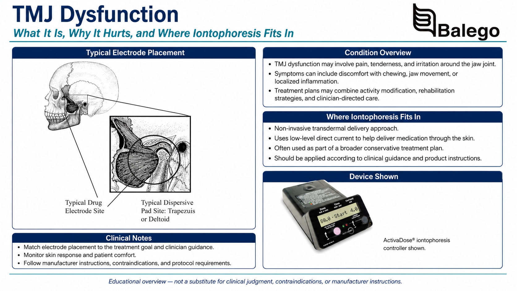
TMJ Iontophoresis at a Glance
| Clinical question | Practical answer |
|---|---|
| Is “TMJ dysfunction” one diagnosis? | No. TMD includes multiple joint, muscle, and headache-related conditions. |
| Which presentation is the most plausible iontophoresis candidate? | Localized, unilateral, joint-dominant pain that can be reproduced over the preauricular TMJ region. |
| Is iontophoresis a primary treatment for muscle-dominant TMD? | Usually not. Exercise, behavioral care, manual therapy, and load modification generally match myalgia more directly. |
| What did the principal adult iontophoresis trial find? | Mandibular function improved in a small group with capsulitis and disc displacement without reduction, but pain did not decrease significantly. |
| Where is the medication electrode positioned? | Over the professionally identified preauricular joint target, anterior to the tragus, while maintaining clearance from the orbit and temporal treatment restrictions. |
| Where is the return electrode placed? | On an appropriate ipsilateral major muscle, commonly the shoulder region, rather than the opposite side of the face or head. |
| Is 40 mA-min always required? | No. It was used in the adult clinical trial, but current dosing must follow the prescription and product-specific limits. |
| What should determine whether treatment continues? | Jaw function, familiar pain, eating tolerance, motion, locking frequency, and skin response. |
First, determine what type of TMD is present.
The phrase “TMJ pain” does not identify the affected structure or mechanism.
| Clinical presentation | Common findings | Likely iontophoresis relevance |
|---|---|---|
| TMJ arthralgia or capsular pain | Familiar pain reproduced by preauricular palpation, opening or lateral movement | May be considered when symptoms are focal and medication has been ordered |
| Disc displacement without reduction, or closed lock | Reduced opening, deviation toward the affected side and a history of locking | Limited adult evidence suggests possible functional benefit in selected patients |
| Disc displacement with reduction | Clicking or popping during movement, sometimes without pain | Painless clicking alone does not require iontophoresis |
| Degenerative joint disease | Crepitus, joint pain, stiffness and possible imaging changes | Requires diagnosis-specific dental or medical management; iontophoresis evidence is limited |
| Masticatory-muscle myalgia | Familiar pain reproduced in the masseter or temporalis rather than directly over the joint | Preauricular joint iontophoresis may not address the primary pain source |
| Headache attributed to TMD | Headache altered by jaw function and reproduced with examination of relevant structures | Requires a broader headache and TMD assessment |
| Cervicogenic or referred pain | Symptoms influenced by neck movement, posture or neurological findings | Treating only the TMJ region is unlikely to address the principal source |
| Dental or oral disease | Tooth pain, infection, abscess, oral lesion or pain with thermal stimuli | Dental evaluation takes priority |
| Persistent multifactorial TMD pain | Symptoms influenced by sleep, stress, widespread pain, fear, mood or sensitization | Emphasize supported self-management, exercise and behavioral approaches |
Iontophoresis is most clinically coherent when the target is localized, superficial, and joint-dominant. It should not be selected merely because pain is felt somewhere near the ear.
Symptoms Consistent With a Temporomandibular Disorder
A patient may report the following:
- Pain in front of one or both ears
- Jaw-muscle pain
- Pain during chewing
- Stiffness or reduced opening
- Painful clicking or popping
- Catching or locking
- Difficulty yawning
- A change in comfortable bite position
- Headache associated with jaw use
- Facial or neck pain
- Difficulty eating firmer food
- Fatigue during prolonged talking
Clicking or popping without pain is common and is not, by itself, an indication for treatment.
Findings Requiring Medical, Dental or Specialist Assessment
A routine rehabilitation or iontophoresis protocol should not delay evaluation when the patient has:
- Recent facial or jaw trauma
- Suspected fracture
- Inability to close the mouth following a yawn, dental procedure or trauma
- Rapidly increasing facial swelling
- Fever, redness or concern for infection
- Dental abscess or severe tooth-related pain
- New facial weakness or sensory loss
- Progressive cranial-nerve findings
- Unexplained weight loss or a suspicious mass
- New severe headache with visual symptoms
- Significant ear drainage, sudden hearing change or acute vestibular symptoms
- A sudden, unexplained change in occlusion
- Juvenile inflammatory arthritis or suspected systemic inflammatory disease
- Severe restriction that continues to worsen
- Pain that cannot be reproduced through a musculoskeletal jaw examination
Jaw, facial, and oral pain may arise from conditions other than TMD, making differential diagnosis essential.
TMD Examination Checklist
A useful examination should identify whether symptoms arise primarily from the joint, muscles, or another region and establish measurable baselines.
1. Clarify the history
Document:
- Exact pain location
- Unilateral or bilateral symptoms
- Sudden or gradual onset
- Duration and progression
- Trauma history
- Jaw locking or catching
- Clicking, popping or crepitus
- Morning versus evening pattern
- Pain during chewing, talking or yawning
- Clenching, bracing or gum-chewing habits
- Dental procedures or orthodontic history
- Headache, ear or cervical symptoms
- Sleep quality
- Widespread pain or other persistent-pain conditions
- Previous dental, medical or rehabilitation treatment
- Current medications and allergies
- Patient priorities and functional goals
Avoid automatically attributing TMD to posture, stress, occlusion, or one behavioral habit. Several physical, psychological, and social factors may interact, particularly when symptoms have become persistent.
2. Identify the patient’s familiar pain
The examination should attempt to reproduce the pain the patient recognizes as the primary complaint.
Ask whether symptoms reproduced during the following:
- TMJ palpation
- Masseter palpation
- Temporalis palpation
- Maximum unassisted opening
- Assisted opening
- Lateral excursion
- Protrusion
- Chewing or biting simulation
are the patient’s familiar symptoms, rather than merely pressure or stretching discomfort.
The Diagnostic Criteria for Temporomandibular Disorders use structured history and examination procedures to improve classification of pain-related TMD.
3. Observe mandibular movement
Observe the opening and closing pathway for:
- Straight movement
- Corrected deviation
- Persistent deviation
- Guarding
- Jerky movement
- Clicking
- Crepitus
- Early or late movement asymmetry
Deviation toward one side does not establish a diagnosis by itself. Interpret it together with pain, motion, locking history, and joint findings.
4. Measure jaw mobility
Measure:
- Pain-free opening
- Maximum unassisted opening
- Maximum assisted opening when appropriate
- Right lateral excursion
- Left lateral excursion
- Protrusion
Record the measurement method so the same procedure can be repeated.
Do not rely on one universal “normal” opening value. Body size, age, pain, joint mechanics, and the measurement technique all affect interpretation.
5. Palpate the joint and chewing muscles
Palpate bilaterally and compare:
- Preauricular TMJ region
- Masseter
- Temporalis
Additional muscle or cervical examination may be appropriate according to the history.
Record:
- Whether familiar pain is reproduced
- Exact pain location
- Joint versus muscle predominance
- Side-to-side differences
- Tenderness intensity
- Referred pain
- Skin condition at a potential electrode site
Aggressive internal palpation or routine testing of every possible muscle is not required for all patients.
6. Evaluate joint sounds carefully
Determine whether a click, pop, or crepitus is
- Painful or painless
- Consistent or intermittent
- Present during opening, closing or both
- Associated with locking
- Associated with functional limitation
Do not treat a sound simply because it is audible.
7. Screen nearby and competing regions
Depending on the presentation, assess:
- Cervical motion
- Upper-quarter neurological status
- Headache pattern
- Ear symptoms
- Dental or oral findings
- Facial sensation
- Cranial-nerve function when clinically indicated
- Generalized joint hypermobility
- Sleep and behavioral contributors
- Psychosocial distress and pain-related disability
A Chvostek test is not a routine diagnostic test for TMD and does not help classify common jaw joint or masticatory muscle disorders.
8. Establish functional baselines
Possible outcomes include the following:
- Jaw Functional Limitation Scale
- Patient-Specific Functional Scale
- Pain during a standardized chewing task
- Maximum comfortable opening
- Time tolerated during conversation
- Number or frequency of locking episodes
- Ability to eat selected food textures
- Headache frequency
- Sleep interruption
- Pain during yawning or dental hygiene
A modality should not be judged solely by tenderness immediately after treatment.
Imaging: When Is It Useful?
Imaging is not automatically necessary for uncomplicated, clinically diagnosed TMD.
It may be considered when there is
- Trauma
- Suspected fracture
- Persistent closed lock
- Concern for degenerative joint disease
- Significant progressive restriction
- Unexplained occlusal change
- Suspected inflammatory arthritis
- A mass or unusual swelling
- Failure to progress despite appropriate care
- A need to clarify internal joint structure before a procedure
MRI is generally the most informative study for disc position and soft-tissue internal derangement. CT or cone-beam CT is more useful for osseous change. Imaging findings must still be interpreted alongside symptoms because structural findings and pain do not always correspond directly.
What Does the Evidence Say About TMJ Iontophoresis
Evidence is limited and applies to narrowly defined populations.
Adult randomized clinical trial
A double-blind randomized trial studied 27 patients with concurrent TMJ capsulitis and disc displacement without reduction. Patients received three treatments every other day. The active-treatment group received dexamethasone sodium phosphate with lidocaine, delivered at 40 mA-min per session. The medication electrode was placed over the involved TMJ, and the return electrode was placed over the ipsilateral trapezius.
The active-treatment group demonstrated the following:
- An average six-millimeter increase in active opening
- An increase in lateral excursion
- Improvement in a mandibular dysfunction index
- No statistically significant reduction in reported pain
The authors concluded that iontophoresis improved mandibular function but did not clearly reduce pain in this selected population. Temporary electrode-site redness and transient dizziness were reported.
Juvenile idiopathic arthritis research
A retrospective pilot study examined dexamethasone iontophoresis for TMJ involvement in children and adolescents with juvenile idiopathic arthritis. The authors reported apparent safety and clinical improvement but specifically called for prospective controlled research. This pediatric inflammatory-arthritis population is fundamentally different from typical adult mechanical or persistent TMD and should not be used to promise comparable results for ordinary jaw pain.
Practical evidence summary
| Question | Evidence-based interpretation |
|---|---|
| Does TMJ iontophoresis reliably reduce pain? | The principal adult trial did not demonstrate significant pain reduction. |
| Can it improve jaw movement? | A small adult trial found improved opening and lateral movement in a specific closed-lock/capsulitis population. |
| Is it proven for muscle-dominant TMD? | No strong condition-specific evidence supports preauricular iontophoresis for masticatory myalgia. |
| Is it equivalent to a steroid injection? | No direct evidence establishes equivalence. |
| Is pediatric JIA evidence applicable to adult TMD? | No. The populations and disease mechanisms differ substantially. |
| Should iontophoresis be used alone? | No. It is best considered as one possible adjunct within a reversible, function-centered plan. |
Where Iontophoresis May Fit
Iontophoresis may be considered when:
- The patient has a reasonably clear joint-dominant diagnosis
- Familiar pain localizes to one preauricular TMJ
- The skin is intact
- A prescriber has ordered an appropriate water-soluble ionized medication
- The treatment objective is specific and measurable
- The electrode can be positioned safely away from the orbital region
- The return electrode can be placed ipsilaterally over an appropriate major muscle
- The patient can remain relatively still during treatment
- A defined reassessment plan has been established
It is less likely to be useful when:
- Pain is predominantly muscular
- Symptoms are widespread or poorly localized
- Painless clicking is the only finding
- A dental, otologic or neurological condition has not been excluded
- Symptoms suggest an acute dislocation or fracture
- The patient has significant altered facial sensation
- The electrode would extend toward the eye
- Safe return-electrode placement cannot be achieved
- Passive treatment is replacing exercise and self-management
- Previous applications produced no meaningful functional change
Iontophoresis Is Not the Same as a TMJ Injection
The original page describes iontophoresis as an alternative to hypodermic corticosteroid injection. That comparison requires qualification.
| Feature | Iontophoresis | Intra-articular injection |
|---|---|---|
| Skin puncture | No | Yes |
| Delivery route | Transdermal medication transport using direct current | Medication placed through a needle into or around a selected joint structure |
| Anatomical precision | Influenced by skin, medication, current, electrode and tissue characteristics | Depends on needle placement and whether imaging guidance is used |
| Evidence for TMD | Limited and diagnosis-specific | Varies by diagnosis, medication and procedure |
| Main risks | Skin irritation, burns, medication reaction and current-related discomfort | Procedural pain, infection, bleeding, tissue injury and medication-related risks |
| Direct equivalence established? | No | No direct injection-versus-iontophoresis equivalence has been established |
Iontophoresis does not guarantee that a medication reaches the same intra-articular location or concentration as an injection. It should be selected on its own clinical rationale rather than presented as a needle-free version of the same procedure.
Comparing Balego Iontophoresis Options for the TMJ Region
The preauricular face is a small, contoured, high-movement treatment area near the eye, ear, and hairline. Product fit and supervision are therefore more important than selecting the largest reservoir or fastest dose.
| Balego option | Potential TMJ advantage | Important facial-placement limitation | Practical role |
|---|---|---|---|
| ActivaDose II Dose Controller with Trivarion Small electrodes | Adjustable current and dose with a smaller, flexible active electrode and direct clinician monitoring | The clinician must confirm that the entire drug matrix fits the preauricular target without approaching the orbit | Often the most practical Balego workflow for closely supervised unilateral TMJ treatment |
| ActivaDose II with Ionto4 Small Square electrodes | The smallest Ionto4 reservoir option and a complete active/return treatment kit | The square adhesive footprint may be less convenient near the tragus, hairline or cheek contour | Alternative controller-based option when the facial footprint fits safely |
| ActivaDose II with buffered iontophoresis electrode kits | Provides several electrode shapes with a buffered return-electrode workflow | Larger shapes may be inappropriate for a small preauricular target | Useful when the clinic already uses this controller-based electrode system |
A controller-based setup offers adjustable current, treatment-time calculation, and active monitoring throughout the session. The ActivaDose II supports prescribed dosing from 0 to 80 mA-min and automatically calculates treatment time based on the selected current and dose. Medication and disposable electrodes are purchased separately.
For the TMJ region, "smallest practical" does not mean "smallest adhesive at any cost." The entire drug matrix must contact intact skin while remaining safely clear of the orbit, external ear opening, hair, and deep skin folds.
Facial Iontophoresis Safety Principles
Balego’s electrode directions list several precautions that are particularly important near the head:
- Do not place the system across the temporal regions
- Do not use it for orbital-region treatment
- Do not apply over damaged or denuded skin
- Do not use in a patient with a cardiac pacemaker or another sensitive implanted device
- Do not use when the patient is sensitive to the prescribed medication
- Remove nearby jewelry
- Do not tape, bind or compress either electrode
- Stop and inspect the site when the patient reports pain
- Limit treatment according to the medication polarity and electrode instructions
What “do not place across the temporal regions” means for a TMJ workflow
Do not create a current path:
- From one temple to the other
- From one preauricular region to the opposite side of the face
- Across the forehead
- Through the orbital region
- Between electrodes placed on opposite sides of the head
A unilateral active electrode with an ipsilateral shoulder-region return electrode is more consistent with the adult trial and avoids a transcranial electrode arrangement.
Step-by-Step TMJ Iontophoresis Protocol
Step 1: Confirm a joint-dominant treatment target
Before preparing the device, document:
- The specific TMD classification or working diagnosis
- Why the joint rather than a chewing muscle is the treatment target
- The exact side
- The familiar pain reproduced over the TMJ
- Baseline jaw motion
- Baseline functional limitation
- The intended role of iontophoresis
A diagnosis of “jaw pain” is insufficient.
Step 2: Define the treatment objective
Examples include:
- Improve comfortable mouth opening
- Improve lateral excursion
- Improve tolerance for chewing
- Reduce joint-related pain that prevents jaw exercise
- Reduce the frequency of function-limiting locking
- Improve tolerance for dental hygiene
- Improve the ability to speak for a required period
The objective should describe a function, not simply “reduce inflammation.”
Step 3: Verify the medication order
Confirm:
- Medication name
- Formulation
- Concentration
- Electrical polarity
- Prescribed dose
- Current limitations
- Treatment frequency
- Medication allergies
- Product compatibility
- Patient-specific precautions
Medication is not included with iontophoresis controllers, electrodes, or patches.
General explanations of polarity, direct current, and mA-min dosing are already available in our Iontophoresis Frequently Asked Questions and should not be duplicated on this condition-specific page.
Step 4: Inspect both electrode sites
Inspect the preauricular region and intended return site for the following:
- Cuts
- Pimples
- Razor irritation
- Dermatitis
- Recent scars
- Abnormal sensation
- Facial hair or sideburn interference
- Makeup, oil or skin products
- Jewelry
- Previous electrode reaction
Do not shave the area immediately before treatment. Do not place the active matrix over the external ear opening or allow the adhesive to approach the eye.
Step 5: Select the active-electrode size
Choose the smallest electrode that:
- Safely holds the prescribed medication volume
- Covers the identified preauricular target
- Maintains complete contact
- Remains outside the orbital region
- Does not bridge the ear
- Does not fold into the hairline
- Does not sit on damaged skin
- Is compatible with the prescribed controller and dose
A larger electrode is not automatically safer or more effective.
Step 6: Prepare the skin without causing irritation
Follow the electrode instructions for use.
Clean and dry the sites sufficiently to remove oil, makeup, and residue, but do not scrub facial skin to the point of redness or abrasion. Any irritation created before direct-current treatment may increase discomfort.
Current electrode directions instruct clinicians to clean the site for approximately six to eight seconds, allow it to dry, and avoid razor nicks, pimples, wounds, and other damaged skin.
Step 7: Prepare the drug electrode off the patient
- Place the electrode on a stable surface
- Add the prescribed medication according to the fill instructions
- Hydrate the entire drug matrix
- Avoid dry spots
- Do not overfill
- Do not underfill
- Do not use a medication suspension when the electrode requires a water-soluble preparation
- Confirm the medication polarity before attaching the lead
Do not fill the electrode while it is attached to the patient.
Step 8: Place the active drug electrode
A typical joint-focused placement is the following:
- Unilateral
- Anterior to the tragus
- Centered over the clinically identified TMJ target
- Positioned so the entire drug matrix contacts intact skin
- Safely clear of the eye and orbital region
- Free from hair, deep creases and the external ear opening
Confirm placement by having the patient gently open and close the mouth before treatment begins. The electrode should remain flat and should not lift, fold, or migrate.
Do not press forcefully on the medication reservoir.
Step 9: Place the return electrode
Position the return electrode over an appropriate ipsilateral major muscle according to the device and electrode instructions.
The adult trial used the ipsilateral trapezius. An appropriate ipsilateral shoulder or upper-arm site may also be considered when compatible with the product instructions and clinical circumstances.
Do not place the return electrode:
- On the opposite side of the face
- On the opposite temple
- Across the forehead
- In the orbital region
- Directly over the neck
- Over jewelry
- Beneath a tight garment or strap
- Where the patient will lie or lean against it
Step 10: Position the patient
Use a supported upright or semi-reclined position.
During controller-based treatment, ask the patient to avoid:
- Talking
- Chewing
- Repeated opening and closing
- Wide yawning
- Turning the head repeatedly
- Touching the electrodes
- Pulling the leads
- Leaning on the return electrode
Movement can disrupt contact and may trigger a controller reject warning.
Step 11: Deliver the prescribed dose
The adult randomized trial used:
- 40 mA-min per treatment
- Current adjusted to tolerance
- A typical session time of approximately 20 minutes
- Three treatments performed every other day
That research protocol should not be converted into a universal treatment rule.
Current dosing must account for:
- Medication order
- Medication polarity
- Electrode limits
- Device limits
- Facial skin sensitivity
- Patient tolerance
- Previous response
Electrode directions specify a maximum of 80 mA-min for negative-polarity delivery and 40 mA-min for positive-polarity delivery for the electrode system described on that page. Different products may have different limits.
Step 12: Monitor continuously
Ask the patient to report the following immediately:
- Burning
- Sharp pain
- Increasing stinging
- Strong itching
- Dizziness
- Visual symptoms
- Ear symptoms
- Metallic or unusual taste
- New numbness
- Electrode movement
- Lead-wire pulling
Pause treatment and inspect the skin and electrode contact when symptoms occur. Discontinue and follow appropriate medical procedures when symptoms do not resolve promptly.
The adult trial reported temporary erythema and transient dizziness, reinforcing the need for active observation during preauricular treatment.
Step 13: Inspect and document
After treatment, record:
- Diagnosis or clinical classification
- Treated side
- Medication
- Concentration
- Polarity
- Dose
- Current
- Treatment time
- Electrode model and size
- Active-electrode placement
- Return-electrode placement
- Patient sensation
- Skin response
- Change in motion
- Change in the selected functional task
- Instructions given
Temporary redness may occur. Blistering, persistent pain, pronounced irritation, swelling, or a medication reaction requires appropriate follow-up.
Pair Iontophoresis With Reversible TMD Care
The National Institute of Dental and Craniofacial Research recommends beginning with simple, reversible measures and avoiding treatment that permanently changes the joint, teeth, or bite unless clearly justified. Initial care may include softer foods, heat or cold, gentle exercise, and reducing jaw-clenching or gum-chewing habits.
Early symptom-management strategies
Depending on the diagnosis:
- Temporarily reduce very chewy or hard foods
- Cut food into smaller pieces
- Avoid gum
- Avoid repeatedly testing the jaw
- Limit exceptionally wide opening
- Support the jaw during a large yawn when helpful
- Reduce daytime clenching or bracing
- Maintain comfortable jaw movement
- Use heat or cold as directed
- Continue normal activity within tolerable limits
A soft-food strategy should generally be temporary. Prolonged avoidance of normal jaw loading can reinforce fear and reduce functional capacity.
Jaw exercise and movement retraining
Exercise should match the diagnosis and irritability.
Possible components include:
- Controlled opening in a comfortable range
- Tongue-supported opening
- Lateral movement
- Protrusion and retrusion control
- Isometric jaw exercise
- Coordination training
- Gradual chewing exposure
- Cervical or postural exercise when examination findings support it
Do not forcefully stretch a recently locked or acutely irritable joint without first confirming the diagnosis.
Manual therapy
Manual therapy may include carefully selected
- Joint mobilization
- Soft-tissue treatment
- Cervical techniques
- Movement retraining
The goal is to improve function and provide a pathway toward self-management, not to create indefinite dependence on passive treatment.
Chronic TMD pain
For adults with chronic TMD pain lasting at least three months, a 2023 BMJ guideline strongly favored interventions including cognitive behavioral therapy with or without selected co-interventions, supported jaw exercise, mobilization, and trigger point approaches. The guideline did not identify passive modalities as the central foundation of long-term care.
Comparing Common TMD Treatment Approaches
| Treatment approach | Primary purpose | Best clinical fit | Important limitation |
|---|---|---|---|
| Education and habit modification | Reduce unnecessary aggravation and improve self-management | Most TMD presentations | Generic advice such as “stop stressing” is inadequate |
| Temporary diet modification | Reduce high-load chewing during an irritable period | Acute joint or muscle pain | Long-term restriction may reduce normal loading and food choice |
| Jaw exercise | Restore motion, control, strength and confidence | Joint- and muscle-related TMD when appropriately dosed | Exercises must match the diagnosis and irritability |
| Manual therapy | Improve movement and reduce symptom barriers | Selected joint, muscle or cervical findings | Benefit should transfer to active self-management |
| Behavioral treatment | Address clenching, pain coping, sleep and persistent-pain contributors | Chronic or multifactorial TMD | Requires appropriate provider access and patient engagement |
| Intraoral appliance | Protect teeth or alter loading temporarily in selected patients | Specific dentist-directed indications | Evidence varies; it should not permanently alter the bite |
| Iontophoresis | Deliver prescribed ionized medication through intact skin | Localized joint-dominant symptoms | Evidence is limited and does not establish reliable pain reduction |
| Injection or arthrocentesis | Provide intra-articular medication or mechanically lavage the joint | Selected arthrogenous disorders after specialist evaluation | Invasive and diagnosis-dependent |
| Surgery | Address severe structural disease in carefully selected cases | Destructive disease or refractory severe dysfunction | Permanent, invasive and not routine first-line TMD care |
Reassessment: What Meaningful Improvement Looks Like
Use the same measures collected before treatment.
Joint symptoms
- Familiar preauricular pain
- Pain during opening
- Pain during lateral excursion
- Joint tenderness
- Locking frequency
- Painful joint noise
- Morning stiffness
Mandibular function
- Pain-free opening
- Maximum unassisted opening
- Assisted opening when indicated
- Right and left excursion
- Protrusion
- Movement pathway
- Ability to close after opening
Daily function
- Chewing selected food textures
- Yawning
- Talking
- Toothbrushing
- Dental care
- Singing
- Instrument performance
- Work communication
- Sleep
- Headache burden
Treatment safety
- Facial skin response
- Shoulder return-electrode response
- Dizziness
- New sensory symptoms
- Ear or visual symptoms
- Medication reaction
Reasons to modify or stop iontophoresis
- No meaningful functional improvement during the defined trial
- Pain worsens
- Opening continues to decrease
- Locking becomes more frequent
- Symptoms become predominantly muscular
- The original joint target no longer reproduces familiar pain
- Repeated skin irritation occurs
- Dizziness or neurological symptoms recur
- Safe electrode placement cannot be maintained
- Passive treatment is delaying active rehabilitation
- Findings suggest another dental, neurological, inflammatory or structural diagnosis
The absence of pain relief does not necessarily mean that movement has not changed, as demonstrated in the adult trial. Both pain and function should be measured.
Documentation Template
TMD classification or working diagnosis: Right/left TMJ __________.
Differential diagnoses considered: __________.
Familiar pain reproduced by: __________.
Baseline mobility: Pain-free opening __________ mm; maximum unassisted opening __________ mm; right excursion __________ mm; left excursion __________ mm.
Functional limitation: Symptoms limit __________.
Reason iontophoresis was selected: Localized joint symptoms interfere with __________.
Medication order: Medication __________; concentration __________; polarity __________.
Delivery system: Controller __________; active electrode __________; return electrode __________.
Placement: Active electrode positioned __________; return electrode positioned __________.
Treatment details: Dose __________ mA-min, current __________ mA, and time ___________.
Patient response: Sensation __________; dizziness/other symptoms __________.
Skin response: Active site __________; return site __________.
Post-treatment findings: Opening __________ mm; functional response __________.
Active rehabilitation plan: __________.
Reassessment criterion: __________.
For billing-oriented support, link clinicians to our Iontophoresis Reimbursement Codes and Documentation Guide.
Frequently Asked Questions
Can iontophoresis help TMJ pain?
It may help selected patients with localized joint-dominant TMD. The principal adult clinical trial found improved mandibular movement and dysfunction scores but did not find significant pain reduction.
Is TMJ the same as TMD?
No. TMJ refers to the temporomandibular joint. TMD refers to disorders involving the joint, chewing muscles, or associated headache conditions.
Which TMD type is most appropriate for iontophoresis?
The most plausible candidate is a localized, unilateral arthrogenous presentation in which familiar pain is reproduced over the TMJ and a suitable medication has been prescribed. Evidence is particularly limited outside capsulitis and disc displacement without reduction.
Where is the medication electrode placed?
The active electrode is generally placed anterior to the tragus over the involved preauricular TMJ target. The entire drug matrix must remain on intact skin and be safely clear of the eye, ear opening, and hairline.
Where is the return electrode placed?
A return electrode may be placed on an appropriate ipsilateral major muscle, such as the shoulder or trapezius region, according to the device and electrode instructions.
Can one electrode be placed over each TMJ?
Do not create an electrode path across the temporal regions or through the head. Bilateral facial electrode placement is not an appropriate default arrangement. Treating one side at a time with an ipsilateral shoulder-region return is more consistent with the available adult trial.
Should the return electrode be placed on the neck?
The neck is not the preferred return site for this protocol. Use an appropriate ipsilateral major muscle that provides full contact and does not create a prohibited current path.
Is 40 mA-min the standard TMJ dose?
The adult randomized trial used 40 mA-min, but this is not a universal prescription. Dose must follow the medication order, polarity, device limits, electrode limits, and patient response.
How many treatments are required?
The adult trial used three treatments every other day. The original Balego page suggested up to six treatments, but neither number should be applied automatically. Establish a defined trial and reassess before continuing.
Is dexamethasone included with the controller or electrodes?
No. Medication is supplied separately and must be selected and prescribed by an appropriately licensed professional.
Is dexamethasone iontophoresis equivalent to a steroid injection?
No. The delivery route, depth, anatomical precision, and evidence differ. No direct evidence establishes that TMJ iontophoresis is equivalent to an intra-articular injection.
Can iontophoresis treat jaw muscle pain?
A preauricular drug electrode targets the joint region, not the full masseter or temporalis muscle. Muscle-dominant TMD generally requires a different treatment rationale.
Can IontoPatch be worn on the face?
A wearable patch should not be treated as the automatic choice for TMJ treatment. The clinician must verify the specific patch instructions, electrode footprint, prescribed dose, and safe clearance from the eye, ear, and temporal region. Closely monitored controller-based treatment is often more practical for this small facial target.
Should clicking be treated?
Painless clicking is common and usually does not require treatment. Painful clicking, locking, or functional limitation warrants a diagnosis-specific examination.
Does an abnormal bite cause TMD?
Current research does not support the general belief that a “bad bite” or orthodontic braces routinely cause TMD. Avoid irreversible bite-changing treatment without a clear and well-supported indication.
When should a patient see a dentist or orofacial pain specialist?
Referral is appropriate when the diagnosis is uncertain, dental disease is suspected, locking persists, occlusion changes unexpectedly, conservative care fails, imaging is needed or an invasive procedure is being considered.
Clinical Takeaway
The essential question is not whether iontophoresis can be placed near the jaw. It is whether the patient has a localized joint-dominant TMD that matches the treatment.
A careful examination should distinguish TMJ arthralgia or capsulitis from muscle pain, painless disc clicking, closed lock, dental disease, cervical referral, and persistent multifactorial pain. Available adult evidence suggests that dexamethasone-and-lidocaine iontophoresis may improve mandibular function in selected patients with capsulitis and disc displacement without reduction, but it should not be promoted as a reliable pain-relief treatment or an equivalent replacement for intra-articular injection.
When used, facial iontophoresis requires conservative unilateral placement, strict avoidance of temporal and orbital current paths, active monitoring, and a clear functional reassessment plan. The treatment should support movement, chewing, communication, and self-management—not replace them.
Explore our iontophoresis controllers, electrodes, and patches, review the Iontophoresis Electrode Directions for Use, or browse additional iontophoresis evaluation and treatment protocols.


