



Calcaneal Bursitis Iontophoresis Protocol: Retrocalcaneal vs. Superficial Heel Bursitis
“Calcaneal bursitis” can refer to two different sources of pain at the back of the heel:
- Retrocalcaneal bursitis, involving the bursa between the Achilles tendon and the upper-back portion of the calcaneus
- Superficial calcaneal bursitis, also called retro-Achilles bursitis, involving tissue between the skin and the back of the Achilles tendon
These conditions can produce pain in nearly the same region, but their anatomy, aggravating factors, and treatment priorities differ. Retrocalcaneal bursitis also frequently overlaps with insertional Achilles tendinopathy and mechanical compression around a prominent posterosuperior calcaneus.
Iontophoresis may be considered when a qualified healthcare professional identifies a localized treatment target and a prescriber orders a compatible ionic medication. It should be used as a selective symptom-modifying adjunct, not as a replacement for footwear changes, compression management, progressive rehabilitation, or evaluation of an accompanying Achilles tendon disorder.
Clinical notice: Medication selection, formulation, concentration, polarity, electrical dose, electrode placement, frequency and treatment duration require an appropriate order and professional judgment. Follow the current instructions for the medication, controller, electrode or wearable patch.
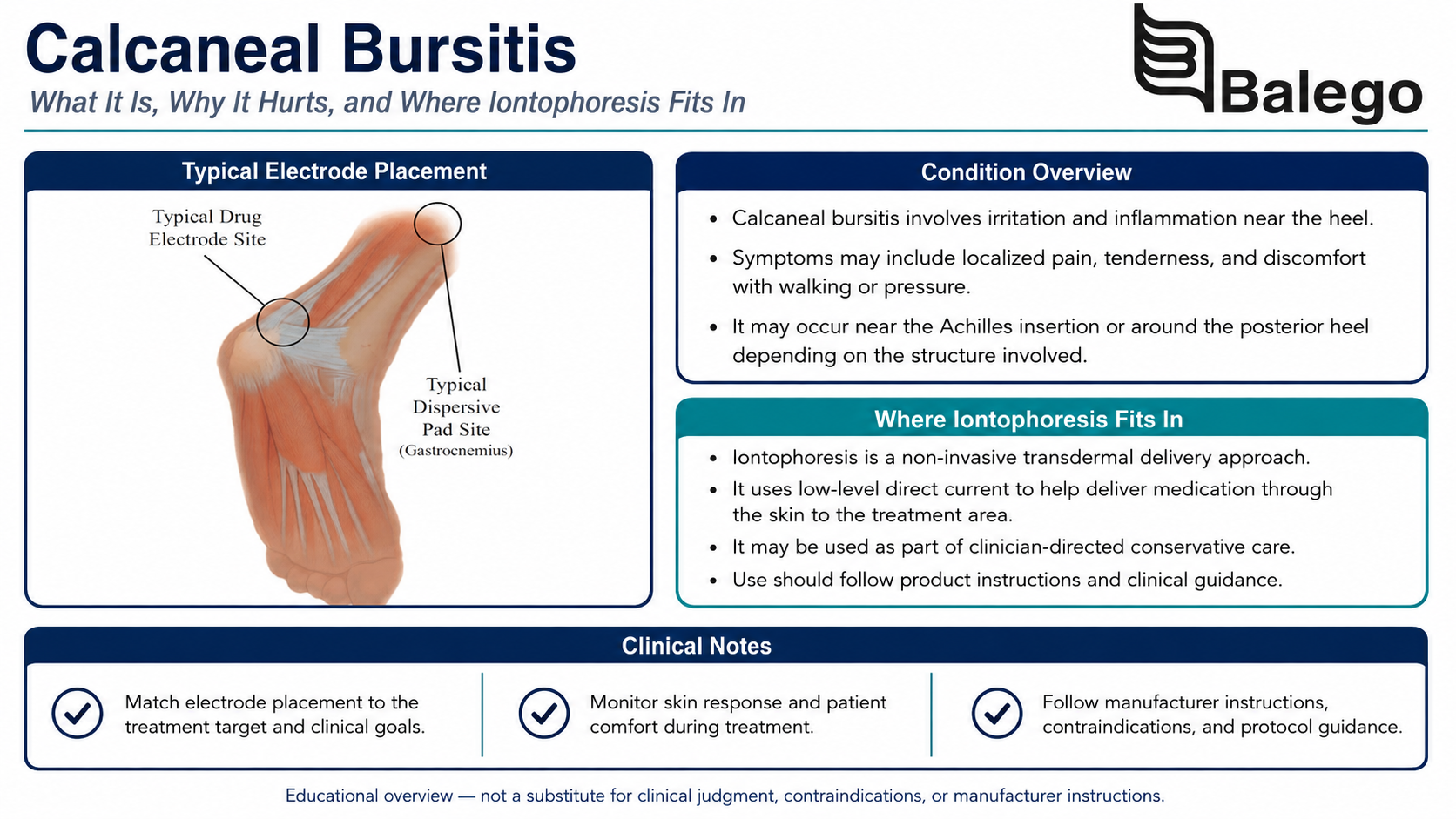
Heel Bursitis at a Glance
| Clinical question | Practical answer |
|---|---|
| Are retrocalcaneal and superficial bursitis the same condition? | No. One lies deep to the Achilles tendon; the other lies between the tendon and skin. |
| What commonly aggravates superficial bursitis? | Direct pressure and friction from a shoe’s heel counter, straps, or rigid footwear. |
| What commonly aggravates retrocalcaneal bursitis? | Compressive loading between the Achilles tendon and calcaneus, especially during deeper dorsiflexion. |
| Does heel bursitis always occur by itself? | No. Retrocalcaneal bursitis may coexist with insertional Achilles tendinopathy or Haglund-related anatomy. |
| Is every painful bump behind the heel bursitis? | No. Tendinopathy, tendon tear, bone prominence, infection, inflammatory disease, and other conditions may look similar. |
| Where may iontophoresis fit? | As a prescribed adjunct for a localized, accessible treatment region with intact skin. |
| Is 40 mA-min the universal dose? | No. Dose must follow the prescription and delivery system instructions. |
| What is usually most important for long-term improvement? | Reducing the provoking compression, correcting footwear conflicts, and gradually restoring calf and Achilles loading capacity. |
Retrocalcaneal vs. Superficial Calcaneal Bursitis
| Feature | Retrocalcaneal bursitis | Superficial calcaneal bursitis |
|---|---|---|
| Location | Between the anterior Achilles tendon and posterosuperior calcaneus | Between the skin and posterior Achilles tendon |
| Depth | Deep | Superficial |
| Typical tenderness | Anterior or immediately beside the distal Achilles insertion | Directly beneath a tender skin-level heel bump |
| Common aggravating factor | Dorsiflexion-related compression and repetitive Achilles loading | Shoe-counter pressure, straps and external friction |
| Visible swelling | May appear beside the Achilles tendon | Often forms a more obvious superficial bump |
| Common overlap | Insertional Achilles tendinopathy or Haglund-related compression | Skin irritation, callus or pressure-related swelling |
| Electrode challenge | The bursa is deep and cannot be directly accessed beneath the tendon with a surface pad | The target is more superficial, but pressure and irritated skin can make adhesion difficult |
| Rehabilitation emphasis | Reduce insertional compression and restore graded calf loading | Remove direct external pressure and allow the superficial tissue to settle |
Symptoms That May Suggest Retrocalcaneal Bursitis
A patient may report the following:
- Deep pain at the back of the heel
- Tenderness immediately anterior to the Achilles tendon
- Pain when the ankle moves into dorsiflexion
- Pain with uphill walking, running or stair climbing
- Discomfort during heel raises
- Swelling beside the distal Achilles tendon
- Morning stiffness
- Symptoms that resemble insertional Achilles tendinopathy
Retrocalcaneal bursitis may arise with repetitive loading, inflammatory or crystalline arthropathy, infection, or mechanical impingement. Achilles insertional abnormalities often coexist, making precise classification important.
Symptoms That May Suggest Superficial Calcaneal Bursitis
A patient may report the following:
- A tender bump directly beneath the skin
- Pain from the back of a shoe
- Redness or irritation over the bump
- Discomfort that improves in open-backed footwear
- Pain from skates, ski boots, work boots or rigid athletic shoes
- Symptoms that are less pronounced when barefoot
- Sensitivity to direct touch rather than deep ankle movement
A rigid or prominent heel counter can compress the soft tissues over the back of the calcaneus. A Haglund-related bony prominence may increase this conflict, but the presence of a prominence on imaging does not by itself establish the pain source.
Calcaneal Bursitis vs. Other Causes of Posterior Heel Pain
| Possible condition | Distinguishing considerations |
|---|---|
| Insertional Achilles tendinopathy | Pain and load sensitivity are concentrated within the distal tendon attachment; tendon thickening or calcification may be present |
| Midportion Achilles tendinopathy | Symptoms generally occur several centimeters above the insertion rather than directly at the heel |
| Partial Achilles tear | May follow a sudden event and produce weakness, bruising or a focal tendon defect |
| Complete Achilles rupture | Sudden pop, impaired push-off, altered resting position or abnormal calf-squeeze response |
| Haglund-related posterior heel pain | A bony prominence may contribute to shoe and tendon compression but is not synonymous with bursitis |
| Posterior ankle impingement | Deeper ankle pain, often aggravated by plantar flexion rather than direct posterior-heel compression |
| Calcaneal stress injury | Focal bone pain, impact intolerance and positive calcaneal compression findings |
| Sural nerve irritation | Burning, tingling or altered sensation along the posterolateral ankle |
| Inflammatory enthesitis | Bilateral symptoms, prolonged stiffness or additional inflammatory joint findings |
| Infection | Progressive redness, warmth, swelling, drainage, fever or systemic illness |
| Gout or crystalline disease | Acute severe swelling and redness with relevant medical history |
| Plantar heel disorder | Pain is concentrated beneath the calcaneus rather than behind the Achilles |
Our separate Achilles Tendinopathy Iontophoresis Protocol addresses tendon-specific rupture screening, loading, and return-to-activity decisions.
Red Flags and Referral Findings
Do not begin a routine heel bursitis protocol when the patient has
- A sudden pop or tearing sensation
- Immediate loss of push-off strength
- Inability to perform a straight-knee heel raise
- Rapid bruising or swelling after an acute injury
- A palpable tendon defect
- Fever or systemic illness
- Rapidly increasing redness or warmth
- Drainage, broken skin or suspected infection
- Severe night pain without a mechanical pattern
- Progressive numbness or weakness
- A suspected calcaneal stress injury
- A major recent increase in pain after corticosteroid injection
- Symptoms suggesting systemic inflammatory disease
- Substantial swelling after trauma
A suspected Achilles rupture, infection, fracture, or inflammatory disease requires medical evaluation rather than routine iontophoresis.
Heel Bursitis Examination Checklist
1. Clarify the symptom history
Document:
- Exact pain location
- Sudden or gradual onset
- Symptom duration
- Recent footwear changes
- Use of boots, skates, orthoses or braces
- Changes in running, walking, jumping or hill exposure
- Morning stiffness
- Pain during and after activity
- Response during the following day
- History of Achilles symptoms
- Prior injections or procedures
- Inflammatory, metabolic or rheumatologic history
- Medication use and allergies
- Patient goals
Ask whether the symptoms improve when the shoe is removed. A strong relationship to direct heel-counter pressure supports superficial involvement but does not exclude an underlying insertional Achilles disorder.
2. Inspect the posterior heel
Compare both sides for:
- Location of swelling
- Skin redness
- Callus or blistering
- A visible posterior prominence
- Swelling beside the Achilles tendon
- Bruising
- Scarring
- Shoe-pressure marks
- Open skin or drainage
- Resting ankle position
3. Map the pain precisely
Ask the patient to point with one finger.
Determine whether the familiar pain is:
- Directly behind the Achilles tendon
- Anterior to the tendon
- At the tendon insertion
- Several centimeters above the heel
- At the posterolateral ankle
- Beneath the calcaneus
- Diffuse or radiating
A precise location is necessary before choosing a medication-electrode site.
4. Palpate the relevant structures
Palpate:
- Superficial skin-level swelling
- Medial and lateral sides of the retrocalcaneal space
- Achilles insertion
- Midportion Achilles tendon
- Posterior calcaneus
- Sural-nerve region
- Adjacent gastrocnemius and soleus
The two-finger squeeze maneuver applies medial and lateral pressure anterior to the Achilles tendon and may reproduce retrocalcaneal-bursa pain. It supports the clinical examination but is not independently diagnostic.
Avoid repeatedly compressing a highly irritable bursa merely to reconfirm that it hurts.
5. Assess ankle movement
Evaluate:
- Weight-bearing dorsiflexion
- Non-weight-bearing dorsiflexion
- Plantar flexion
- Inversion and eversion
- Symptom response to deeper dorsiflexion
- Side-to-side differences
Deep dorsiflexion can increase compression around the Achilles insertion and retrocalcaneal region. The finding should be interpreted alongside tendon loading, palpation, and footwear response.
6. Assess calf loading capacity
Depending on irritability and tendon integrity, assess the following:
- Bilateral heel raise
- Single-leg heel raise
- Heel-raise height
- Number of controlled repetitions
- Bent-knee calf loading
- Walking push-off
- Stair climbing
- Running or jumping when appropriate
Reduced capacity may reflect pain, an accompanying insertional tendon disorder, or deconditioning.
7. Examine footwear
Inspect:
- Heel-counter rigidity
- Location of seams
- Shoe collar height
- Fit around the posterior heel
- Evidence of rubbing
- Difference between symptomatic and comfortable shoes
- Space available for a heel lift
- Whether an orthosis raises the heel into a more irritating part of the shoe
- Whether the shoe is too loose and repeatedly slides
“Change shoes” is not sufficiently specific. Identify exactly which part of the shoe conflicts with the painful structure.
8. Establish functional baselines
Possible measures include:
- Foot and Ankle Ability Measure
- Lower Extremity Functional Scale
- Patient-Specific Functional Scale
- Walking duration
- Stair tolerance
- Shoe-wearing tolerance
- Heel-raise repetitions
- Pain during a standardized dorsiflexion or calf-loading task
- Running distance
- Work-shift tolerance
When Is Imaging Useful?
Routine imaging is not necessary for every superficial, footwear-related presentation.
Consider radiographs, ultrasound, or MRI when:
- The diagnosis remains uncertain
- An Achilles tear is suspected
- Symptoms persist despite appropriate management
- A prominent posterior calcaneal process may affect treatment
- There is substantial tendon thickening
- Inflammatory disease is suspected
- A stress injury is possible
- Surgery or another invasive procedure is being considered
Radiographs help evaluate calcaneal shape, spurs, and other osseous findings. Ultrasound can assess the Achilles tendon, retrocalcaneal bursa, and surrounding soft tissue dynamically. MRI may help evaluate the complete tendon-bursa-bone complex when a more extensive insertional disorder is suspected.
What Does the Evidence Say About Iontophoresis?
The most directly relevant recent research was a 2025 retrospective study involving 159 patients diagnosed with insertional Achilles tendinopathy or retrocalcaneal bursitis. Eighty-seven patients received at least three dexamethasone-iontophoresis treatments.
The researchers found:
- No significant between-group differences in pain
- No significant between-group differences in range of motion
- Larger posterior heel spurs among patients receiving more treatments
- A nonsignificant tendency toward surgery among those with larger spur or Haglund measurements
- A possible short-term role for dexamethasone iontophoresis, without evidence of significant long-term functional improvement
The authors called for prospective research. Because the study was retrospective and combined insertional tendinopathy with retrocalcaneal bursitis, it does not establish an optimal dose, treatment frequency, or universal benefit for heel bursitis.
Responsible interpretation
| Question | Evidence-based answer |
|---|---|
| Is iontophoresis proven to resolve heel bursitis? | No |
| Is it established as superior to rehabilitation alone? | No |
| Does recent research show a clear long-term pain or motion benefit? | No significant difference was found |
| May it provide temporary symptom modification? | Possibly, but certainty is limited |
| Is 40 mA-min the established calcaneal bursitis dose? | No |
| Are three to six every-other-day sessions proven? | No |
| Should treatment address footwear and loading? | Yes |
| Should insertional Achilles involvement be assessed? | Yes |
Iontophoresis Is Not Equivalent to a Bursa Injection
The original Balego page characterizes iontophoresis as a needle-free alternative to corticosteroid injection. The comparison needs qualification.
| Feature | Iontophoresis | Image-guided bursa injection |
|---|---|---|
| Skin puncture | No | Yes |
| Delivery route | Medication transported through intact skin using direct current | Medication placed through a needle into the selected bursa |
| Anatomical precision | Influenced by skin, electrode position, medication and tissue depth | Guided by needle position, often with ultrasound or fluoroscopy |
| Ability to reach the deep bursa | Surface treatment cannot be assumed to reproduce an intra-bursal injection | Medication can be placed directly within the bursa |
| Evidence | Limited and largely indirect | Procedure-specific evidence exists but risks and indications differ |
| Principal risks | Skin irritation, burn and medication reactions | Infection, bleeding, tendon exposure and other procedural or medication risks |
| Direct equivalence established? | No | No |
Iontophoresis may be selected because it is noninvasive, but it should not be described as producing the same anatomical delivery or clinical effect as an injection.
When Iontophoresis May Fit the Plan
Iontophoresis may be considered when:
- The diagnosis is reasonably clear
- Symptoms are focal
- The skin is intact
- A medication has been appropriately ordered
- The medication’s polarity is verified
- The target can be covered without excessive bony pressure
- Pain is preventing participation in rehabilitation
- The patient can avoid shoe or surface pressure over the electrode
- A measurable functional objective is established
- The plan includes footwear and activity changes
It is a weak match when:
- The skin is blistered, infected or broken
- Pain is diffuse
- An Achilles rupture is suspected
- The condition is primarily caused by ongoing direct shoe pressure that has not been changed
- The deep target cannot be reasonably localized
- The electrode will be compressed by a shoe or brace
- Symptoms are predominantly neurological
- A systemic inflammatory disorder has not been evaluated
- Repeated applications have not improved function
Step-by-Step Calcaneal Bursitis Iontophoresis Protocol
Step 1: Classify the posterior heel presentation
Document whether the working diagnosis is:
- Retrocalcaneal bursitis
- Superficial calcaneal bursitis
- Insertional Achilles tendinopathy with bursal involvement
- Haglund-related posterior heel compression
- Another condition requiring a different pathway
Do not use “heel bursitis” as the sole treatment justification.
Step 2: Define the functional objective
Examples include:
- Improve tolerance for normal footwear
- Improve walking tolerance
- Improve ability to perform introductory calf loading
- Reduce pain during stairs
- Improve tolerance for work boots
- Support a graded return to running
“Reduce inflammation” is not a complete functional objective.
Step 3: Verify the medication order
Confirm:
- Medication name
- Formulation
- Concentration
- Ionic polarity
- Prescribed dose
- Medication volume
- Frequency
- Allergies
- Patient-specific precautions
- Compatibility with the delivery system
Medication is not included with Balego iontophoresis controllers, electrode kits, or wearable patches.
For general medication polarity and mA-min guidance, link to our Iontophoresis Frequently Asked Questions rather than repeating the complete information here.
Step 4: Inspect the active and return sites
Check for:
- Cuts
- Blisters
- Abrasions
- Rash
- Folliculitis
- Drainage
- Infection
- Altered sensation
- Bruising
- Severe swelling
- Previous electrode reaction
- Shoe-related skin damage
Skin-preparation instructions differ among product families. Controller-based electrode kits may include alcohol prep pads, while IontoPatch instructions specify soap and water rather than alcohol. Follow the exact instructions for the product being used.
Step 5: Select an electrode that fits the heel contour
The posterior heel is narrow, curved, and exposed to external pressure.
Choose an electrode that:
- Covers the localized target
- Holds the prescribed medication volume
- Maintains full contact
- Does not bridge a deep crease
- Does not wrap excessively around the ankle
- Avoids unnecessary pressure over the calcaneus
- Can remain free from footwear
- Does not cover broken or highly irritated skin
A butterfly or flexible electrode may conform better than a large square electrode around the Achilles region.
Step 6: Prepare the medication electrode
For a controller-based system:
- Prepare the drug electrode off the patient
- Use the prescribed medication volume
- Saturate the full medication matrix as directed
- Avoid dry areas
- Avoid overfilling
- Verify polarity before connecting the lead
- Follow the electrode’s current and dose limits
Use our Iontophoresis Electrode Directions for Use for the complete setup and safety instructions.
Step 7: Position the medication electrode
Retrocalcaneal target
The bursa lies deep to the Achilles tendon, so a surface electrode cannot be placed directly inside or beneath it. The clinician should place the medication matrix over the intact skin that best overlies the localized symptomatic region while recognizing the depth limitation.
Placement principles include the following:
- Use examination findings or imaging to localize the side and level
- Avoid pressing the electrode firmly into the Achilles-calcaneal interval
- Avoid placing a large rigid electrode directly over a prominent calcaneus
- Confirm that the patient’s familiar pain is deep rather than purely skin-level
- Keep footwear off the electrode
- Stop if new burning, numbness or radiating symptoms develop
Superficial calcaneal target
When the superficial bursa is the selected target:
- Place the medication matrix over the focal swelling only when the skin is intact
- Avoid blistered, abraded or infected skin
- Ensure the electrode lies flat rather than tenting over the bump
- Avoid a shoe, strap or brace over the electrode
- Reconsider iontophoresis when the swelling is too prominent for reliable contact
Step 8: Position the return electrode
Place the return electrode over appropriate intact muscle tissue according to the product instructions.
The calf may be practical when it provides the following:
- Complete contact
- Adequate separation
- No pressure from the treatment table
- No compression from clothing
- Minimal lead-wire tension
- Normal skin sensation
Do not bind or tightly tape either electrode.
Step 9: Position the patient
Prone or side-lying positioning with the foot free from table pressure may help protect the active electrode.
Confirm that:
- The heel is not resting on the electrode
- The patient is not repeatedly moving the ankle
- Lead wires remain slack
- Both sites can be inspected
- Clothing does not press against either electrode
- The patient can immediately report discomfort
Step 10: Deliver the prescribed dose
Do not automatically use the previous page’s:
- 40 mA-min dose
- Every-other-day schedule
- Three-to-six-treatment course
- Lower dose based only on the description “sensitive skin”
The appropriate treatment depends on:
- Medication order
- Polarity
- Electrode limits
- Controller or patch limits
- Skin condition
- Target depth and size
- Patient response
- Functional reassessment
The ActivaDose II Iontophoresis Dose Controller permits selection of a prescribed dose and current while displaying treatment time and monitoring delivery. Its available range does not override lower medication or electrode limits.
Step 11: Monitor the patient
Ask the patient to report:
- Burning
- Sharp pain
- Increasing stinging
- Strong itching
- New numbness
- Electrode movement
- Pressure against the heel
- Lead-wire pulling
- Unexpected ankle or Achilles symptoms
Pause and inspect the skin and setup when discomfort develops. Do not merely reduce the current without determining the cause.
Step 12: Inspect and document
Record:
- Working diagnosis
- Treated side
- Medication
- Concentration
- Polarity
- Dose
- Current
- Treatment time
- Electrode or patch model
- Active placement
- Return placement
- Skin response
- Patient sensation
- Immediate functional response
- Footwear instructions
- Rehabilitation plan
- Decision to continue, modify or discontinue treatment
Comparing Iontophoresis Options for Posterior Heel Placement
| Balego option | Potential heel advantage | Important limitation | Practical fit |
|---|---|---|---|
| ActivaDose II with Trivarion electrodes | Flexible Small and Butterfly options are designed for curved and uneven locations, including the Achilles region | Requires a controller, lead wires and protected positioning | Clinics prioritizing contour, current control and direct monitoring |
| ActivaDose II with Ionto4 electrodes | Butterfly and several square sizes support different footprints and medication volumes | A large square may not conform to the narrow heel | Clinics wanting standardized single-treatment kits |
| ActivaDose II with buffered iontophoresis electrode kits | Provides multiple active-electrode shapes and a buffered return electrode | Electrode size must be matched carefully to the heel contour | Clinics preferring Ag/AgCl drug electrodes and buffered return pads |
| IontoPATCH wearable patches | Self-contained treatment without external wires | Shoes, socks, walking, perspiration and the narrow heel contour may compromise fit | Selected patients who can protect the application site throughout wear |
| ACTIVApatch wearable patches | No-saline wearable models offer several treatment durations | Wireless design does not remove the problems of footwear pressure and heel movement | Clinics seeking simplified wearable preparation when site and medication are compatible |
Trivarion’s form-fitting design and butterfly configuration are specifically intended for curved areas such as the Achilles region. Wearable patches remove the controller and lead wires but may be more difficult to protect from shoe pressure and repeated ankle motion.
For full wearable-system specifications, use our IontoPatch vs. ACTIVApatch comparison.
Remove the Provoking Compression
Superficial bursitis
The first priority is usually eliminating direct external pressure.
Consider:
- Temporarily using open-backed footwear
- Choosing a softer heel counter
- Avoiding seams that contact the swelling
- Adjusting lacing to reduce heel movement
- Using different work or sport footwear
- Avoiding tight straps
- Checking whether an insert raises the heel into an irritating shoe edge
A patient cannot be expected to improve while the same rigid shoe repeatedly compresses the superficial bursa.
Retrocalcaneal bursitis
Reduce internal compression around the Achilles insertion when it reproduces symptoms.
Temporary modifications may include:
- Limiting deep ankle dorsiflexion
- Reducing steep uphill activity
- Avoiding heel drops below a step
- Reducing running or jumping volume
- Performing heel raises from a level surface
- Reviewing posterior shoe pressure
- Using a clinician-directed heel lift when appropriate
A recent randomized trial involving sport-active patients with insertional Achilles tendinopathy found better outcomes from a rehabilitation program that reduced tendon compression through education, heel lifts, and controlled dorsiflexion during progressive exercise. Although the study addressed insertional tendinopathy rather than isolated bursitis, its compression-management principles may be relevant when the two conditions coexist.
Heel Lifts: Temporary Load-Management Tools
A heel lift can reduce ankle dorsiflexion and may temporarily decrease compression around the insertional Achilles-retrocalcaneal region. It should be selected according to the patient, shoe, and diagnosis rather than automatically prescribed.
Balego options include:
- Clearly Adjustable Heel Lift, adjustable in 1 mm increments
- Balego Adjustable Heel Lift, using removable layers
- Our full Heel Lifts, wedges and Inserts Collection
| Heel-lift consideration | Why it matters |
|---|---|
| Height | Excessive elevation may create a new pressure point |
| Shoe depth | The heel must remain secure inside the shoe |
| Heel-counter position | Raising the heel may move the painful region against a different shoe edge |
| Bilateral vs. unilateral use | Depends on gait, comfort and clinician judgment |
| Duration | Often used as a temporary tool rather than a permanent substitute for rehabilitation |
| Gradual removal | A sudden reduction in lift height may rapidly increase dorsiflexion and tendon demand |
Short-term research in insertional Achilles tendinopathy suggests that heel lifts can improve symptoms and gait measures, but they remain one component of a broader program.
Progressive Rehabilitation
Stage 1: Settle the provoking irritation
Depending on the presentation:
- Remove direct shoe pressure
- Reduce deeper dorsiflexion
- Reduce running, jumping or hill exposure
- Maintain tolerable walking and conditioning
- Avoid repeatedly compressing the bursa to “test” it
- Avoid aggressive friction massage over an inflamed superficial bursa
- Maintain comfortable ankle movement
Stage 2: Restore calf loading
Begin with tolerable options such as:
- Seated heel raises
- Supported bilateral heel raises
- Isometric plantar-flexion work
- Heel raises from a level surface
- Controlled bent-knee calf loading
Do not begin an insertional presentation with unsupported heel drops below a step when deep dorsiflexion reproduces symptoms.
Stage 3: Progress strength
Advance through:
- Single-leg heel raises
- External resistance
- Knee-straight calf strengthening
- Knee-bent soleus strengthening
- Step-ups
- Walking inclines
- Progressive occupational or sport tasks
Stage 4: Reintroduce compression and energy-storage demands
When basic loading is tolerated, gradually restore:
- Greater dorsiflexion range
- Hills
- Faster walking
- Running
- Pogo hops
- Jumping
- Acceleration
- Sport-specific footwear
The progression should reflect the patient’s response during activity and over the following day.
Comparing Common Heel-Bursitis Treatments
| Treatment | Primary purpose | Best fit | Important limitation |
|---|---|---|---|
| Footwear modification | Remove direct external pressure | Especially important for superficial bursitis | Does not restore calf capacity |
| Activity-load modification | Reduce the mechanical demand exceeding current tolerance | Retrocalcaneal or insertional overlap | Must be followed by graded return |
| Progressive calf loading | Restore Achilles and calf function | Tendon involvement or deconditioning | Requires appropriate range and dosage |
| Heel lift | Temporarily reduce dorsiflexion and insertional compression | Selected retrocalcaneal or insertional presentations | Can create new shoe-pressure problems |
| Iontophoresis | Deliver prescribed ionic medication through intact skin | Localized symptoms with suitable skin and professional order | Long-term effectiveness remains uncertain |
| Cold therapy | Temporary symptom management | Irritable, swollen presentations | Does not address footwear or loading |
| Injection | Directly place medication into a selected bursa | Selected medically evaluated cases | Invasive and anatomically distinct from iontophoresis |
| Shockwave therapy | Symptom-focused mechanical treatment | Some chronic insertional presentations | Evidence and tolerance vary |
| Surgery | Address refractory mechanical or tendon pathology | Persistent cases after comprehensive nonsurgical care | Operative risks and rehabilitation |
Reassessment: What Meaningful Improvement Looks Like
Symptoms
- Reduced posterior-heel tenderness
- Reduced swelling
- Less pain from footwear
- Reduced pain during dorsiflexion
- Improved morning comfort
- Reduced next-day reaction
Capacity
- Improved heel-raise tolerance
- Improved calf strength
- Improved walking distance
- Improved stair tolerance
- Increased running or jumping tolerance
- Improved shoe tolerance
Skin and treatment response
- Acceptable electrode-site response
- No blistering
- No spreading rash
- No medication reaction
- No worsening skin breakdown
Reasons to reconsider the plan
- No meaningful functional improvement
- Increasing swelling
- New bruising or weakness
- New neurological symptoms
- Repeated electrode irritation
- Ongoing shoe pressure has not been corrected
- The pain localizes more clearly to the Achilles tendon
- Symptoms become systemic or bilateral
- A tendon tear, infection or stress injury becomes more likely
- Passive treatment is delaying progressive rehabilitation
Documentation Template
Clinical classification: Right/left retrocalcaneal bursitis, superficial calcaneal bursitis, or mixed insertional Achilles-bursal presentation.
Differential diagnoses considered: __________.
Red-flag and tendon-integrity screening: __________.
Focal treatment target: __________.
Footwear finding: __________.
Baseline functional measure: Walking/shoe tolerance/heel raises/FAAM/LEFS/PSFS __________.
Reason iontophoresis was selected: Localized symptoms interfere with __________.
Medication order: Medication __________; formulation __________; concentration __________; polarity __________.
Delivery system: Controller/patch __________; electrode model and size __________.
Placement: Medication electrode __________; return electrode __________.
Treatment details: Dose __________ mA-min, current __________ mA, and time ___________.
Patient and skin response: __________.
Immediate functional response: __________.
Footwear and loading plan: __________.
Reassessment criterion: __________.
For documentation and payer-oriented information, direct clinicians to our Iontophoresis Reimbursement Codes and Documentation Guide.
Frequently Asked Questions
What is calcaneal bursitis?
It is irritation of a bursa near the posterior heel. The two principal posterior bursae are the deep retrocalcaneal bursa and the superficial bursa between the Achilles tendon and skin.
Is retrocalcaneal bursitis the same as Achilles tendinopathy?
No. They involve different structures, but they frequently coexist and can produce similar symptoms.
Is superficial calcaneal bursitis caused by shoes?
Direct pressure from a rigid heel counter or strap is a common aggravating factor. The footwear conflict must be corrected for the tissue to settle.
Is Haglund deformity the same as heel bursitis?
No. Haglund deformity describes a bony prominence. It may contribute to soft-tissue compression, but a prominence does not automatically mean the bursa or tendon is symptomatic.
Where does retrocalcaneal bursitis hurt?
Pain is usually deep at the back of the heel, immediately anterior to or beside the distal Achilles tendon.
Where does superficial calcaneal bursitis hurt?
Pain and swelling are closer to the skin, directly where footwear presses on the back of the heel.
Can iontophoresis help calcaneal bursitis?
It may be considered a prescribed symptom-modifying adjunct. Recent retrospective research did not demonstrate significant long-term pain or range-of-motion differences for insertional Achilles or retrocalcaneal bursitis patients receiving multiple dexamethasone-iontophoresis treatments.
Is iontophoresis equivalent to a steroid injection?
No. Iontophoresis delivers medication through the skin, while an injection places medication through a needle into a selected anatomical location. Their depth, precision, risks, and evidence differ.
Where should the medication electrode be placed?
Place it over the intact skin that best overlies the clinically identified target. Retrocalcaneal and superficial bursitis require different positioning.
Can the electrode be placed directly on a blistered heel?
No. Do not apply over broken, infected, or substantially irritated skin.
Where should the return electrode be placed?
For controller-based treatment, an appropriate calf-muscle location may be used when it provides full contact and complies with the electrode and controller instructions.
Is 40 mA-min the standard dose for heel bursitis?
No. The previous Balego page used 40 mA-min, but current evidence does not establish it as a universal calcaneal-bursitis dose.
How many treatments are required?
There is no universal course. Continue only when a defined treatment trial produces meaningful functional improvement with acceptable skin tolerance.
Can a shoe be worn over an iontophoresis patch?
Avoid direct shoe pressure over the patch. Compression can disturb adhesion, press on the reservoirs, and worsen the original irritation.
Can a patient walk with a heel patch?
Possibly, when permitted by the clinician and product instructions. Walking must not fold, compress, or loosen the patch.
Should the calf be stretched?
Stretching should be selected carefully. Deep dorsiflexion may aggravate retrocalcaneal or insertional compression, particularly during an irritable stage.
Should heel raises be performed below a step?
Not initially when deeper dorsiflexion reproduces insertional or retrocalcaneal symptoms. Begin from a level surface and progress range gradually.
Can heel lifts help?
A clinician-directed heel lift may temporarily reduce dorsiflexion and compression, but it must fit the shoe without creating a new heel-counter pressure point.
When is imaging appropriate?
Imaging may be useful when the diagnosis is uncertain, a tendon tear or stress injury is suspected, symptoms persist, inflammatory disease is possible, or a procedure is being considered.
When does posterior heel pain require urgent evaluation?
Prompt assessment is appropriate after a sudden pop with weakness, extensive bruising, suspected rupture, fever, spreading redness, drainage, substantial trauma, or progressive neurological symptoms.
Clinical Takeaway
A calcaneal-bursitis protocol should begin by determining which bursa is involved and whether the Achilles tendon is also part of the presentation.
Superficial bursitis is commonly dominated by external shoe pressure. Retrocalcaneal bursitis is deeper and frequently overlaps with insertional Achilles compression. These presentations should not receive identical electrode placement, footwear advice, or exercise progression.
Iontophoresis may provide a noninvasive medication-delivery option for a carefully selected patient, but it is not anatomically equivalent to an injection and does not remove a bony prominence, alter footwear, or restore calf capacity. Recent retrospective evidence does not show a significant long-term pain or range-of-motion advantage in insertional Achilles and retrocalcaneal bursitis patients.
Long-term treatment should focus on:
- Removing the provoking shoe or surface pressure
- Controlling excessive insertional compression
- Gradually restoring calf and Achilles loading
- Reintroducing dorsiflexion, running and sport demand
- Reassessing the diagnosis when progress stalls
Explore our iontophoresis patches, electrodes, and dose controllers, compare heel lifts and inserts, or browse additional iontophoresis treatment protocols.


