



Hamstring tendon pain can occur near the sitting bone, behind the knee, or along the transition between muscle and tendon. These locations should not be grouped into one generic “hamstring tendonitis” diagnosis.
The first clinical decision is determining whether the patient has:
- Proximal hamstring tendinopathy at the ischial tuberosity
- Distal medial hamstring tendinopathy near the posteromedial knee
- Distal biceps femoris tendinopathy near the posterolateral knee
- An acute muscle strain, partial tear, or tendon avulsion
- Referred pain from the spine, hip, pelvis, or nervous system
This distinction is especially important when iontophoresis is being considered. A localized distal tendon close to the skin presents a different drug-delivery target from the deep proximal hamstring origin beneath the gluteus maximus.
Iontophoresis may be considered when a qualified healthcare professional has identified a focal treatment target and a prescriber has ordered an appropriate ionized medication. It should be described as a selective symptom-modifying adjunct, not as the foundation of hamstring rehabilitation.
Current reviews continue to identify education, load modification, and exercise-based rehabilitation as the cornerstone of nonoperative proximal hamstring treatment. Evidence is still insufficient to recommend one intervention as universally superior.
Clinical notice: Medication selection, concentration, polarity, dose, current, electrode placement, treatment frequency, and activity progression require an appropriate order, professional judgment, and compliance with the medication, electrode, patch, and dose-controller instructions.
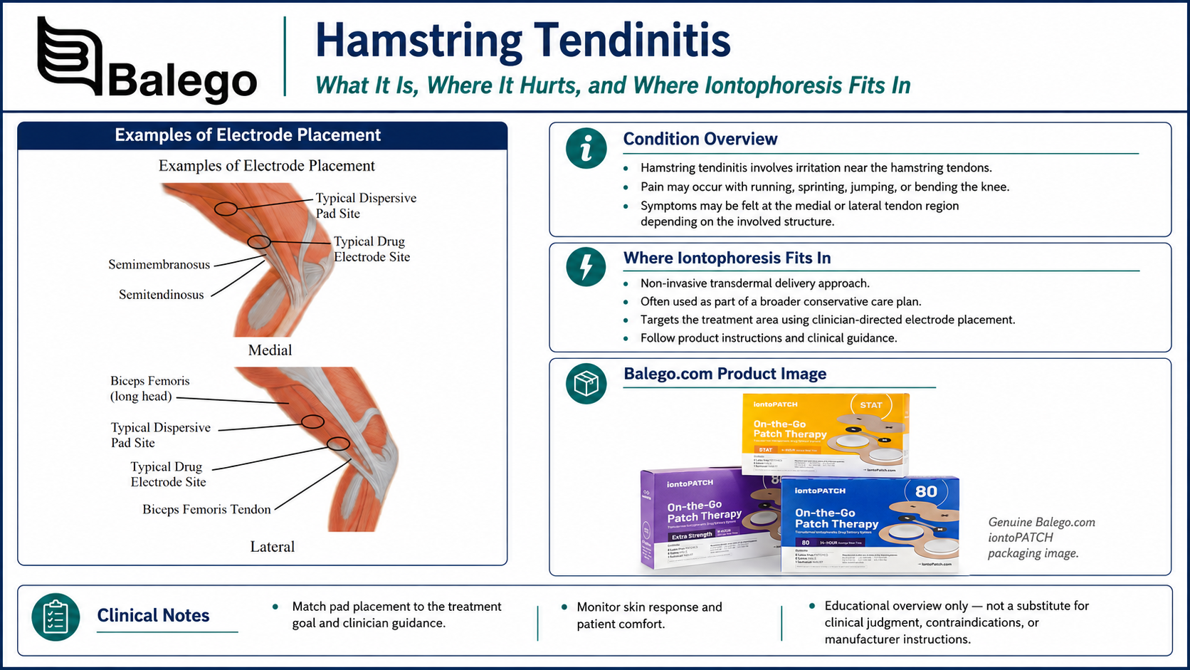
Hamstring Iontophoresis at a Glance
| Clinical question | Practical answer |
|---|---|
| Is every posterior-thigh complaint hamstring tendinopathy? | No. Acute strains, tendon tears, avulsions, nerve-related pain, hip disorders, and lumbar referral may produce similar symptoms. |
| What is the most common proximal presentation? | Localized lower-buttock pain aggravated by sitting, faster running, hills, lunging, or deeper hip flexion. |
| What is a typical distal medial presentation? | Localized posteromedial knee pain associated with the semimembranosus or other medial hamstring structures. |
| What is a typical distal lateral presentation? | Localized pain near the distal biceps femoris tendon and posterolateral knee. |
| Which sites are easiest to target with a surface electrode? | Superficial, well-localized distal tendon sites are generally more accessible than the deep proximal origin. |
| Is there strong hamstring-specific iontophoresis evidence? | No. Direct clinical evidence is sparse, and controlled proximal hamstring studies have evaluated other interventions. |
| Is 40 mA-min the required dose? | No. The current Balego page’s 40 mA-min example is not established as a universal hamstring dose. |
| What is the primary rehabilitation treatment? | Education, activity-load management, and progressive hamstring and kinetic-chain loading. |
| What should determine whether treatment continues? | Functional improvement, load tolerance, strength, symptom response, and acceptable skin tolerance. |
“Hamstring Tendonitis” vs. Hamstring Tendinopathy
The search term "hamstring tendonitis" remains common, but "tendinopathy" is generally more accurate for persistent load-related tendon pain.
The suffix “-itis” implies that inflammation is the primary condition. Persistent tendinopathy can also involve altered tendon structure, reduced mechanical capacity, pain sensitization, compression intolerance, and impaired function.
This distinction changes the treatment emphasis. Passive anti-inflammatory approaches may reduce a temporary symptom barrier, but they do not restore the tendon’s ability to tolerate sitting, running, lifting, sprinting, jumping, or occupational load.
Where Is the Pain? Proximal vs. Distal Hamstring Tendinopathy
| Presentation | Typical pain location | Common aggravating activities | Electrode-placement implication |
|---|---|---|---|
| Proximal hamstring tendinopathy | At or just below the ischial tuberosity—the “sitting bone” | Prolonged sitting, faster running, hills, deep squats, lunges, deadlifts, kicking, and hamstring stretching | The tendon origin is deep and exposed to compression from sitting; surface targeting is less straightforward |
| Distal semimembranosus tendinopathy | Posteromedial knee, often near the tendon’s tibial attachments | Running, cutting, repeated knee flexion, and resisted hamstring loading | A smaller or tendon-oriented electrode may fit a localized superficial target |
| Distal semitendinosus-region pain | Medial knee near the pes anserine region | Repetitive running, cutting, stairs, or resisted knee flexion | Differentiate tendon pain from pes anserine bursitis and medial knee pathology |
| Distal biceps femoris tendinopathy | Posterolateral knee near the fibular-head region | Running, acceleration, deceleration, kicking, and resisted knee flexion | Requires precise localization rather than broad placement over the lateral knee |
| Muscle or myotendinous strain | Posterior thigh, commonly away from the tendon attachment | Sudden sprinting, reaching, kicking, slipping, or rapid eccentric loading | A strain or tear follows a different evaluation and rehabilitation pathway |
| Tendon avulsion | Proximal buttock with sudden severe pain, bruising, and weakness | Sudden forced hip flexion with the knee extending | Requires timely medical assessment rather than routine iontophoresis |
Proximal hamstring tendinopathy commonly produces localized ischial pain that interferes with sitting and physical activity. Higher-speed running, hills, and activities combining hamstring load with hip flexion can be especially provocative because the tendon is both loaded and compressed near the ischial tuberosity.
Distal semimembranosus tendinopathy is much less extensively studied. It is one possible cause of posteromedial knee pain among several competing diagnoses, including meniscal disorders, osteoarthritis, pes anserine pathology, popliteal cysts, gastrocnemius injury, and ligament injury.
Do Not Confuse Tendinopathy With an Acute Hamstring Injury
Although a previous injury may contribute to later tendon symptoms, an acute strain, tear, or avulsion should not be treated as interchangeable with gradual-onset tendinopathy.
| Feature | Tendinopathy | Acute strain or tear |
|---|---|---|
| Onset | Usually gradual or associated with a progressive increase in load | Usually sudden |
| Reported sensation | Ache, stiffness, or localized load-related pain | Sharp pain, tearing, or a pop |
| Bruising | Usually absent | May appear after a more significant injury |
| Weakness | Often load-dependent or related to pain inhibition | May be immediate and substantial |
| Walking | Frequently possible | May be altered or difficult |
| Sitting pain | Common in proximal tendinopathy | May occur after a proximal tear but is not diagnostic |
| Palpable defect | Not expected | May be present in a severe tear or avulsion |
| Initial priority | Classification, load management, and progressive rehabilitation | Determine injury severity and whether imaging or surgical consultation is required |
Red Flags and Findings Requiring Medical Assessment
Do not proceed with a routine iontophoresis protocol when the patient reports or demonstrates:
- A sudden pop or tearing sensation
- Immediate severe weakness
- Rapid bruising or swelling
- A palpable defect
- Inability to walk normally after a significant injury
- Markedly reduced knee-flexion or hip-extension strength
- A suspected proximal tendon avulsion
- Acute ischial pain in a skeletally immature athlete
- Progressive leg numbness or neurological weakness
- Significant back pain with neurological findings
- Fever, redness, unusual warmth, or suspected infection
- Unexplained night pain, systemic illness, or a mass
- Symptoms inconsistent with a load-related tendon presentation
Proximal hamstring injuries exist on a spectrum from tendinopathy and partial tears to complete avulsions. Severe avulsions and some substantial myotendinous injuries may require surgical consultation, whereas most tendinopathy presentations begin with nonoperative management.
Differential Diagnosis of Proximal Hamstring Pain
Lower-buttock pain is not specific to the hamstring origin.
| Possible condition | Distinguishing considerations |
|---|---|
| Lumbar referral or radiculopathy | Back symptoms, neurological findings, diffuse pain, or symptoms altered by spinal movement |
| Sciatic-nerve involvement | Tingling, burning, numbness, radiating pain, or symptoms influenced by neural-tension testing |
| Deep gluteal syndrome | Buttock and neural symptoms influenced by hip position or prolonged sitting |
| Ischiofemoral impingement | Deep buttock or groin-region symptoms influenced by hip position |
| Hip-joint disorder | Restricted hip motion, groin pain, mechanical symptoms, or pain reproduced by hip testing |
| Sacroiliac or pelvic referral | Symptoms reproduced by regional examination rather than isolated tendon load |
| Ischiogluteal bursitis | Direct sitting pain and focal bursal tenderness that may mimic tendon pain |
| Pelvic or sacral stress injury | Load-related bone pain, impact intolerance, or risk factors for bone stress injury |
| Inflammatory enthesitis | Morning stiffness, spinal symptoms, bilateral involvement, or other inflammatory features |
| Proximal hamstring tear | Traumatic onset, weakness, bruising, or imaging-confirmed tendon disruption |
A 2024 review emphasizes that the diagnosis should combine the history, palpation, and active or provocative tests rather than relying on palpation alone. It also notes that lumbar, hip, sacroiliac, sciatic, ischiofemoral, and stress-related conditions may produce similar buttock symptoms.
Differential Diagnosis of Distal Hamstring Pain
Posteromedial knee symptoms
Consider:
- Semimembranosus tendinopathy
- Semitendinosus or pes anserine pathology
- Medial meniscal injury
- Medial-compartment osteoarthritis
- Medial collateral ligament injury
- Popliteal or Baker’s cyst
- Medial gastrocnemius injury
- Popliteus injury
- Referred or neurological pain
Posterolateral knee symptoms
Consider:
- Distal biceps femoris tendinopathy
- Distal biceps femoris tear
- Proximal tibiofibular-joint disorder
- Lateral meniscal pathology
- Posterolateral-corner injury
- Lateral collateral ligament injury
- Popliteus disorder
- Common fibular-nerve irritation
Distal hamstring disorders are less common and less extensively researched than proximal tendinopathy. A broad diagnosis of “hamstring tendonitis” should not be assigned to every posteromedial or posterolateral knee complaint.
Hamstring Tendinopathy Examination Checklist
1. Clarify the history
Document:
- Exact pain location
- Gradual or sudden onset
- Duration and progression
- Previous hamstring injury
- Sitting tolerance
- Running speed and distance
- Hills, sprinting, jumping, and cutting exposure
- Changes in training or occupational load
- Deep-squat, lunge, deadlift, or gardening tolerance
- Pain during and after exercise
- Symptoms during the following 24 hours
- Neurological symptoms
- Lumbar, hip, or knee symptoms
- Medication use and allergies
- Relevant systemic or inflammatory conditions
- Patient goals
For proximal tendinopathy, asking about sitting is especially important. Direct pressure from a hard surface and prolonged hip flexion can increase compression at the hamstring origin.
2. Map the pain precisely
Ask the patient to indicate the primary pain with one finger.
Determine whether the pain is:
- At the ischial tuberosity
- Slightly below the buttock
- In the posterior thigh
- At the posteromedial knee
- At the posterolateral knee
- Diffuse or radiating
- Associated with tingling or numbness
A highly localized tendon complaint is a more plausible iontophoresis target than broad posterior-thigh or radiating pain.
3. Inspect for acute injury findings
Assess for:
- Bruising
- Swelling
- Altered gait
- Visible asymmetry
- A tendon or muscle defect
- Protective movement
- Inability to sit normally
- Skin suitability at a potential treatment site
4. Palpate the suspected tendon
For proximal symptoms, palpation should cover the ischial attachment systematically rather than pressing one generic point.
The proximal tendon footprint is broad and deep beneath the gluteus maximus. The sciatic nerve lies close to the lateral portion of the ischial attachment, making precise diagnosis and cautious treatment planning important.
For distal symptoms, palpate the clinically identified tendon rather than pressing indiscriminately in the central space behind the knee.
5. Assess resisted loading
Depending on the suspected location, assess the following:
- Knee flexion at different angles
- Hip extension
- Long-lever bridge
- Short-lever bridge
- Prone hamstring curl
- Standing hamstring curl
- Single-leg bridge
- Functional hip-hinge tasks
Record:
- Familiar pain location
- Force output
- Side-to-side difference
- Pain versus true weakness
- Response at different hip and knee angles
Proximal hamstring pain during resisted knee flexion may become more apparent as hip flexion increases.
6. Use proximal pain-provocation tests selectively
Common tests include:
- Puranen–Orava test
- Bent-knee stretch test
- Modified bent-knee stretch test
One study reported useful diagnostic properties for these tests in athletes with chronic proximal hamstring symptoms. However, the evidence comes from a limited population, and the tests should be interpreted alongside the history, palpation, loading response, and regional examination.
Aggressive stretching should not be repeated simply to “confirm” the pain, especially when compression sensitivity is high.
7. Assess the kinetic chain
When clinically relevant, evaluate:
- Gluteus maximus strength
- Calf strength
- Quadriceps strength
- Hip control
- Lumbopelvic control
- Running or walking technique
- Overstriding
- Trunk position
- Single-leg control
- Acceleration and deceleration mechanics
Deficits should be treated when demonstrated in the individual—not assumed from the diagnosis.
8. Establish baseline outcome measures
For proximal hamstring tendinopathy, useful measures may include:
- VISA-H
- Patient-Specific Functional Scale
- Sitting tolerance
- Pain during a standardized bridge or knee-flexion task
- Pain during a Puranen–Orava or bent-knee stretch test
- Knee-flexion and hip-extension strength
- Running distance or speed
- Occupational lifting tolerance
The VISA-H was developed to assess symptoms, function, and sporting activity in proximal hamstring tendinopathy.
For distal symptoms, use location-appropriate functional tests such as resisted knee flexion, running, stairs, cutting, or a work-specific activity.
Is Imaging Necessary?
Proximal hamstring tendinopathy is commonly diagnosed from the patient interview and physical examination. Imaging may be appropriate when:
- The diagnosis remains uncertain
- A partial or complete tear is suspected
- Symptoms began traumatically
- Weakness is substantial
- An avulsion is possible
- Neurological symptoms are present
- Progress has stalled despite appropriate rehabilitation
- A procedure or surgical consultation is being considered
MRI is useful for differential diagnosis and is more sensitive than ultrasound for some proximal hamstring abnormalities. Imaging findings must still be matched to the clinical presentation because proximal hamstring changes are also found in people without symptoms.
What Does the Evidence Say About Hamstring Iontophoresis?
Direct hamstring evidence is limited
A systematic review of proximal hamstring tendinopathy included 12 studies involving 424 participants. The reviewed interventions included shockwave therapy, exercise or multimodal rehabilitation, platelet-rich plasma, autologous blood, corticosteroid injection, and surgery. It found insufficient evidence to recommend one treatment over another. Iontophoresis was not among the controlled proximal hamstring interventions evaluated.
A literature review of distal semimembranosus tendinopathy listed iontophoresis among historically used physical-therapy modalities, but the condition itself was supported primarily by small case series and reports rather than high-quality iontophoresis trials.
Accordingly, hamstring iontophoresis should not be described as an evidence-proven protocol comparable with the condition-specific trials available for some other tendons.
Why Proximal Placement Requires Additional Caution
The proximal hamstring origin is
- Deep beneath the gluteus maximus
- Adjacent to the ischial tuberosity
- Exposed to direct sitting pressure
- Near the sciatic nerve
- Frequently aggravated by compression as well as tensile load
A surface electrode over the buttock does not provide the anatomical precision of an image-guided injection and should not be described as delivering medication directly into every portion of the tendon origin.
Because sitting commonly aggravates proximal tendinopathy, a wearable patch that is compressed beneath the patient may be uncomfortable, displaced, or difficult to monitor. The clinician should determine whether the anatomy and treatment objective make surface iontophoresis reasonable before selecting the product.
When Iontophoresis May Be Considered
A clinician may consider iontophoresis when:
- The diagnosis is reasonably clear
- The symptoms are focal
- The skin is intact
- The target can be reached with stable surface contact
- A medication has been appropriately ordered
- Pain is limiting a specific rehabilitation activity
- A meaningful reassessment measure has been selected
- The patient can avoid pressure, friction, and excessive movement over the electrodes
- The treatment is being combined with active rehabilitation
It is less likely to be useful when:
- Pain is diffuse
- Symptoms are primarily neurological
- An acute tear or avulsion is suspected
- The target is too deep or poorly localized
- Sitting will compress a proximal patch
- Clothing or movement will disrupt electrode contact
- The skin is damaged or insensate
- The patient expects passive treatment to replace loading
- Previous applications produced no meaningful functional change
Comparing Iontophoresis Options for Hamstring Sites
This comparison focuses on hamstring-specific fit. Complete dose, fill-volume, and wear-time information remains on the linked product pages.
| Balego option | Potential hamstring advantage | Important limitation | Practical fit |
|---|---|---|---|
| ActivaDose II Controller with IOGEL electrodes | Adjustable controller-based delivery with tendon-oriented elliptical electrode options | Requires lead wires, stable positioning, and clinician monitoring | Localized distal tendon targets or clinics preferring direct control |
| ActivaDose II with Trivarion electrodes | Flexible, form-fitting shapes can help on curved or uneven anatomy | A flexible electrode does not overcome the depth of the proximal hamstring origin | Distal knee contours or other locations where a flat square electrode lifts |
| ActivaDose II with Ionto4 electrodes | Small, medium, large, and butterfly options permit selection by target size and medication volume | Larger sizes should not be used merely because the hamstring is a large muscle group | Clinics wanting several standardized electrode footprints |
| ActivaDose II with buffered iontophoresis electrode kits | Multiple active-electrode shapes combined with a buffered return electrode | Correct anatomical localization remains essential | Controller-based clinics that prefer a buffered return-electrode workflow |
| IontoPATCH wearable patches | Self-contained and wire-free | Sitting, clothing, sweating, hip movement, and knee flexion may compress or loosen the patch | Selected patients who can protect a suitable superficial site |
| ACTIVApatch wearable patches | Several self-contained treatment-time formats | A shorter treatment does not correct poor site access or pressure from sitting | Wireless workflows when the prescribed model and treatment site are compatible |
The ActivaDose II provides clinician-controlled current and dose through compatible wired electrodes. Trivarion emphasizes conformity to curved or uneven anatomy, while Ionto4's and Balego’s buffered kits offer multiple square and butterfly footprints. IontoPATCH and ACTIVApatch provide self-contained wearable options without an external controller.
For full patch-dose and treatment-time comparisons, use our IontoPATCH vs. ACTIVApatch guide instead of reproducing those specifications here.
Step-by-Step Hamstring Iontophoresis Protocol
Step 1: Classify the location and presentation
Document whether the treatment target is:
- Proximal hamstring origin
- Distal semimembranosus
- Distal semitendinosus-region tendon
- Distal biceps femoris
- Another clearly identified superficial structure
Confirm that the presentation is tendinopathy rather than the following:
- Acute muscle strain
- Partial tear
- Tendon avulsion
- Bursitis
- Knee-joint pathology
- Lumbar referral
- Sciatic-nerve involvement
Step 2: Define the functional objective
Examples include:
- Improve tolerance for introductory hamstring strengthening
- Reduce pain that limits normal sitting
- Improve tolerance for walking uphill
- Improve pain during controlled knee-flexion exercise
- Improve participation in a return-to-running program
- Reduce a focal symptom barrier during work activity
“Reduce inflammation” is not a complete functional treatment objective.
Step 3: Verify the medication order
Confirm:
- Medication
- Formulation and concentration
- Electrical polarity
- Prescribed dose
- Treatment frequency
- Medication allergies
- Patient-specific precautions
- Compatibility with the chosen patch or electrode
Medication is not included with most Balego iontophoresis patches, electrodes, or controllers.
For general explanations of polarity and mA-min dosing, link to our Iontophoresis Frequently Asked Questions rather than duplicating that information here.
Step 4: Inspect both electrode sites
Assess for:
- Cuts
- Rash
- Folliculitis
- Bruising
- Recent scar tissue
- Altered sensation
- Significant body hair
- Previous skin reaction
- Pressure from clothing
- Moisture or perspiration
- Areas that will be compressed during sitting or lying
Do not apply the system over damaged, denuded, infected, or substantially insensate skin.
Step 5: Select the electrode size and shape
Choose an electrode that:
- Covers the focal target
- Holds the prescribed medication volume
- Maintains full contact
- Does not fold into a skin crease
- Is not unnecessarily large
- Will not be compressed by sitting, clothing, or a brace
- Matches the contour of the selected site
A large electrode covering the posterior thigh does not compensate for an uncertain diagnosis.
Step 6: Prepare the skin and medication electrodes.
Follow the exact instructions for the selected product.
General controller-based principles include:
- Remove oils and residue
- Clip excessive hair when necessary
- Avoid shaving immediately before treatment
- Prepare the medication electrode off the patient
- Hydrate the entire drug matrix
- Avoid dry areas
- Do not overfill or underfill
- Verify polarity before connecting the lead wires
Our Iontophoresis Electrode Directions for Use contains the complete preparation, filling, polarity, dose limit, and skin-monitoring instructions.
Step 7: Position the active medication electrode
Proximal hamstring target
If proximal iontophoresis is clinically selected:
- Position the drug matrix over the localized ischial treatment target
- Confirm that the placement reproduces the known region rather than diffuse buttock pain
- Avoid positioning that will be compressed by sitting
- Ensure the electrode does not fold beneath the gluteal crease
- Confirm that clothing does not pull or roll the adhesive
- Recognize that the tendon origin is deep and close to the sciatic nerve
- Stop if the patient develops radiating, tingling, or neurological symptoms
The patient should not sit directly on the active electrode during treatment.
Distal medial target
For a confirmed distal medial tendon target:
- Position the drug matrix over the localized tendon site
- Differentiate the tendon from pes anserine, meniscal, bursal, and joint-line pain
- Avoid blind central placement within the popliteal space
- Confirm full contact during a small amount of knee movement
- Reposition if the electrode bridges a crease or lifts
Distal lateral target
For a confirmed distal biceps femoris target:
- Localize the tendon rather than broadly covering the lateral knee
- Confirm that the drug matrix remains on intact soft tissue
- Avoid excessive pressure over a bony prominence
- Stop if stimulation or placement produces new radiating or tingling symptoms
- Check that the electrode remains secure with gentle knee motion
Step 8: Position the return electrode
For a controller-based system, place the return electrode over appropriate intact tissue according to the device and electrode instructions.
Possible regions may include an ipsilateral
- Posterior thigh
- Anterior or lateral thigh
- Calf muscle
The return site should provide:
- Full electrode contact
- Adequate separation
- No pressure from the treatment table
- No tight clothing or compression
- No lead-wire tension
- Intact sensation and skin
Our electrode directions recommend placing the dispersive electrode over a major muscle and prohibit binding or compressing the electrodes.
Step 9: Position the patient
For a proximal target, side-lying or prone positioning may avoid direct pressure on the active electrode.
For a distal target, position the knee comfortably so that:
- The electrode is visible or accessible
- The patient does not repeatedly flex and extend the knee
- The lead wires are not pulled
- Neither electrode is compressed
- The patient can report discomfort immediately
Before starting, test a small amount of normal movement to confirm that the electrode remains flat.
Step 10: Deliver the prescribed dose
Do not automatically use the current page’s 40 mA-min. setting.
The correct treatment depends on:
- The prescription
- Medication polarity
- Medication formulation
- Electrode limits
- Controller or patch limits
- Skin response
- Patient tolerance
- Treatment-site anatomy
The ActivaDose II allows a clinician to select the prescribed dose and current while monitoring treatment time and resistance. Its overall range does not override lower limits imposed by the medication or connected electrode.
Step 11: Monitor continuously
Ask the patient to report:
- Burning
- Sharp pain
- Increasing stinging
- Strong itching
- Electrode movement
- Pressure from the table or clothing
- New tingling or numbness
- Radiating symptoms
- Unexpected weakness
- Lead-wire pulling
Pause treatment and inspect the skin, electrode contact, and equipment when discomfort develops. Do not simply lower the current without investigating the cause.
Step 12: Inspect, document, and reassess
Record:
- Clinical classification
- Treatment side
- Medication and concentration
- Polarity
- Dose
- Current
- Treatment time
- Electrode or patch model
- Active-electrode location
- Return-electrode location
- Patient sensation
- Skin response
- Change in the baseline loading test
- Instructions provided
Temporary redness may occur with direct-current treatment. Blistering, persistent pain, substantial irritation, swelling, or an unusual rash requires appropriate follow-up.
Progressive Rehabilitation Is the Primary Treatment
Iontophoresis does not restore:
- Hamstring force
- Tendon stiffness
- Running tolerance
- Sprinting capacity
- Hip-flexion tolerance
- Kinetic-chain coordination
- Energy-storage and release capacity
Those outcomes require a progressive exercise program matched to the patient’s presentation and goals.
A 2025 clinical commentary proposes a staged proximal hamstring program incorporating isometric exercise, isotonic strengthening, kinetic-chain training, graded compressive loading, and energy-storage activity. The authors emphasize individualized progression rather than a fixed exercise list. (PubMed Central (PMC))
Stage 1: Modify the Most Provocative Load
For proximal symptoms, early modifications may include:
- Reducing faster running
- Temporarily limiting hills
- Modifying deep squats and lunges
- Reducing deep-range deadlifts
- Avoiding prolonged hamstring stretching
- Changing sitting position
- Using a softer sitting surface
- Adjusting chair height and pelvic position
- Reducing long uninterrupted sitting periods
Complete rest is generally not the objective. Less provocative conditioning and strengthening can often continue.
For distal symptoms, modify the activity that reproduces the localized tendon pain, such as:
- Repeated acceleration
- Cutting
- High-volume running
- Kicking
- Repeated knee-flexion exercise
- Deep-loaded knee positions
- Repetitive occupational bending
Stage 2: Introduce Tolerable Hamstring Loading
Possible early exercises include:
- Double-leg bridge
- Single-leg bridge
- Prone hamstring curl
- Standing hamstring curl
- Short-range hip-extension exercise
- Clinician-directed isometric knee flexion
Exercise selection should minimize excessive hip flexion when proximal compression remains highly irritable.
A published staged program uses controlled isometric and isotonic loading, progressing from higher-repetition work to heavier strength loading as tolerated.
Stage 3: Develop Hamstring and Kinetic-Chain Strength
Progressive loading may include:
- Loaded bridges
- Hamstring curls
- Hip thrusts
- Calf raises
- Step-ups
- Knee-extension strengthening
- Hip-abductor or lateral-control exercise
- Lumbopelvic-control training when a deficit is demonstrated
The purpose is to improve the entire lower limb’s capacity to generate, absorb, and distribute force—not to blame one weak muscle for the tendon condition.
Stage 4: Reintroduce Hip-Flexion and Compressive Load
For proximal tendinopathy, gradually restore tolerance for:
- Hip hinging
- Deadlifting
- Lunging
- Deeper squatting
- Picking objects up from the floor
- Gardening
- Occupational lifting
- Sport-specific flexed-hip positions
A current staged model progresses the hip-flexion angle gradually after the patient has demonstrated tolerance for lower-compression strengthening.
Do not begin with aggressive long-range hamstring stretching when compression at the ischial origin is highly provocative.
Stage 5: Restore Energy-Storage and Speed Capacity
Later progression may include:
- Running drills
- Faster running
- Hill exposure
- Acceleration
- Deceleration
- Skipping
- Jumping
- Bounding
- Cutting
- Kicking
- Sport-specific drills
The tendon must tolerate both load magnitude and loading speed. Slow strengthening alone does not establish readiness for sprinting or field sport.
Example Pain-Monitoring Approach
A published proximal hamstring program permits some pain during controlled exercise, commonly using a ceiling around 4/10. It recommends reducing load, repetitions, range, or hip-flexion angle when a repeatable task worsens by more than approximately two points for longer than 24 hours. This is one clinician-directed model, not a universal rule.
The monitoring approach should account for:
- Symptoms during exercise
- Symptoms later that day
- The following morning
- Sitting tolerance
- Performance on a repeatable loading task
- Overall training load
Comparing Hamstring Tendinopathy Treatment Approaches
| Treatment | Main purpose | Evidence position | Important limitation |
|---|---|---|---|
| Education and load management | Reduce excessive compression or loading while maintaining appropriate activity | Considered first-line | Advice must be individualized rather than simply "rest." |
| Progressive exercise | Restore hamstring and kinetic-chain capacity | Cornerstone of nonoperative management | Requires time, adherence, and progression |
| Iontophoresis | Provide localized prescribed medication through intact skin | Hamstring-specific evidence is sparse | Does not restore strength or tendon capacity |
| Shockwave therapy | Apply mechanical impulses to the symptomatic region | Limited evidence; one small trial favored shockwave over a weak comparison program | Cost, discomfort, and uncertainty about ideal selection |
| Corticosteroid injection | Short-term symptom modification in selected cases | Case-series evidence suggests benefits may not persist | Invasive and may have implications for tendon health |
| PRP or autologous blood | Attempt to influence tendon recovery | Low- or very-low-certainty evidence | Invasive, costly, and not clearly superior |
| Surgery | Address selected severe or refractory tendon pathologies. | Used selectively for avulsion or persistent cases | Operative risks and substantial rehabilitation |
The 2021 systematic review rated the available evidence for proximal hamstring interventions as low or very low. A 2024 review similarly recommends education and targeted exercise as first-line management and notes that corticosteroid benefits reported in case series were generally short-term.
Reassessment: What Meaningful Improvement Looks Like
Proximal symptoms
- Increased sitting tolerance
- Reduced localized ischial pain
- Improved tolerance for hills or faster walking
- Improved bridge or knee-flexion loading
- Improved hip-flexion tolerance
- Improved running speed or distance
- Reduced next-day symptom escalation
Distal symptoms
- Reduced posteromedial or posterolateral tenderness
- Improved resisted knee-flexion tolerance
- Improved stair or walking tolerance
- Improved running and cutting
- Reduced pain during acceleration or kicking
- Improved strength in relevant knee positions
Capacity
- Improved knee-flexion strength
- Improved hip-extension strength
- Better single-leg control
- Increased exercise resistance
- Improved repeated-effort tolerance
- Improved speed and energy-storage capacity
- Improved confidence under load
Reasons to reconsider iontophoresis
- No measurable functional change during the defined trial
- Repeated skin irritation
- Increasing weakness
- New bruising or swelling
- New neurological symptoms
- Pain becomes diffuse
- The original tendon is no longer the primary suspected source
- Electrode contact cannot be maintained
- Sitting or clothing repeatedly compresses the treatment site
- Passive care is delaying loading progression
- Findings suggest a tear, avulsion, or alternative diagnosis
Documentation Template
Clinical classification: Right/left proximal hamstring tendinopathy / distal medial hamstring tendinopathy / distal biceps femoris tendinopathy.
Differential diagnoses considered: __________.
Acute tear or avulsion screening: __________.
Focal treatment target: __________.
Baseline provocation test: Test __________; load/range __________; pain __________.
Baseline outcome measure: VISA-H / Patient-Specific Functional Scale / strength / sitting tolerance / other __________.
Functional limitation: Symptoms limit __________.
Reason iontophoresis was selected: Localized symptoms interfere with __________.
Medication order: Medication __________; concentration __________; polarity __________.
Delivery system: Controller/patch __________; electrode model and size __________.
Placement: Medication electrode __________; return electrode __________.
Treatment details: Dose __________ mA-min, current __________ mA, and time ___________.
Patient and skin response: __________.
Immediate functional response: __________.
Loading plan: __________.
Progression or reassessment criterion: __________.
For payer-focused documentation information, direct clinicians to our Iontophoresis Reimbursement Codes and Documentation Guide.
Frequently Asked Questions
Can iontophoresis help hamstring tendonitis?
It may be considered as a localized symptom-modifying adjunct when a clinician has identified a focal tendon target and an appropriate medication has been ordered. Direct hamstring-specific clinical evidence is limited, so iontophoresis should be combined with progressive rehabilitation rather than promoted as a stand-alone treatment.
Is hamstring tendonitis the same as hamstring tendinopathy?
The terms are often used interchangeably in searches. Tendinopathy is generally the more accurate term for persistent load-related tendon pain because symptoms are not necessarily caused by acute inflammation alone.
What is high hamstring tendinopathy?
High or proximal hamstring tendinopathy affects the tendon origin near the ischial tuberosity at the lower buttock. Sitting, faster running, hills, lunging, and deeper hip-flexion activities commonly provoke symptoms.
Where does distal hamstring tendinopathy hurt?
Distal medial symptoms may occur behind and toward the inside of the knee, while distal biceps femoris symptoms are generally more posterolateral. These areas contain several structures, so the diagnosis should be confirmed before treatment.
Where should the medication electrode be placed?
Place the drug matrix over the focal tendon target identified during the clinical examination. Proximal, distal medial, and distal lateral symptoms require different placement.
Can the electrode be placed anywhere along the back of the thigh?
No. Broad posterior-thigh placement is unlikely to be useful when the treatment target has not been identified. Muscle strain, nerve pain, and referred pain require different clinical reasoning.
Is proximal hamstring iontophoresis difficult to perform?
It can be. The proximal tendon origin is deep beneath the gluteus maximus and exposed to compression during sitting. A surface electrode may also be difficult to protect from clothing and movement.
Can a patient sit on a proximal hamstring patch?
Direct pressure should be avoided. Sitting on the patch can compress the reservoir, alter adhesion, increase friction, and prevent skin monitoring.
Should a wearable patch be used for every hamstring condition?
No. Wearable patches may be practical for selected superficial targets, but prolonged sitting, hip motion, knee flexion, perspiration, and clothing can interfere with treatment.
Is dexamethasone included with the electrode or patch?
No. Medication is generally purchased separately and must be selected and ordered by an appropriately licensed professional.
Is 40 mA-min the standard hamstring dose?
No. 40 mA-min, but direct hamstring research has not established that dose as a universal standard. Follow the prescription and product-specific instructions.
How many treatments are required?
There is no evidence-based universal course for hamstring tendinopathy. Establish a defined treatment trial and continue only when reassessment demonstrates meaningful benefit.
Can iontophoresis replace strengthening?
No. It does not restore hamstring strength, tendon-loading capacity, sprint tolerance, or kinetic-chain function.
Should the hamstring be stretched?
Aggressive stretching can increase tensile and compressive load at the proximal tendon origin. Stretching should be selected and progressed according to the diagnosis and irritability rather than automatically prescribed.
Should a patient stop running completely?
Not necessarily. Running may be reduced in speed, volume, hills, or frequency while lower-load conditioning and strengthening continue. More irritable cases may require a temporary interruption.
What exercises are used for proximal hamstring tendinopathy?
Programs may progress from bridges or hamstring curls to heavier isotonic strengthening, kinetic-chain exercise, hip-flexion loading, and eventually faster running and sport-specific drills.
Is a Nordic hamstring curl always required?
No. Nordic curls are one high-load exercise option. Exercise selection should reflect the patient’s current capacity, symptom location, goals, and required activity.
When is imaging appropriate?
Imaging may be helpful after trauma, when a tear or avulsion is suspected, when substantial weakness is present, when the diagnosis remains uncertain, or when progress has stalled despite appropriate care.
When should a patient be referred urgently?
Prompt evaluation is appropriate after a sudden pop with severe weakness, extensive bruising, a palpable defect, difficulty walking, suspected tendon avulsion, or progressive neurological symptoms.
Clinical Takeaway
A hamstring iontophoresis protocol should never begin with the assumption that all posterior-thigh or posterior-knee pain represents the same condition.
The clinician must first distinguish the following:
- Proximal tendinopathy from acute tendon injury
- Tendon pain from muscle strain
- Proximal tendon pain from lumbar, hip, pelvic, or neural referral
- Distal medial tendon pain from other posteromedial knee disorders
- Distal biceps femoris pain from posterolateral knee pathology
Iontophoresis has limited direct evidence for hamstring tendinopathy. It may be considered when the target is localized, the skin is intact, an appropriate medication has been prescribed, and treatment has a measurable functional purpose. It should not be described as equivalent to an injection or as a substitute for rehabilitation.
Long-term recovery depends on restoring the patient’s ability to tolerate compression, hamstring loading, hip flexion, running speed, occupational demands, and sport-specific energy storage.
Explore our iontophoresis patches, electrodes, and dose controllers, review the Iontophoresis Electrode Directions for Use, or browse additional iontophoresis evaluation and treatment protocols.


